Figures & data
Figure 1 Cell-penetrating peptide T9(dR) or TP condensed siRNA and formed nanoparticles with siRNA. (A) Gel retardation assay and (B) size of nanoparticles at 25°C and 37°C. **P-values <0.05 were considered to be significant.
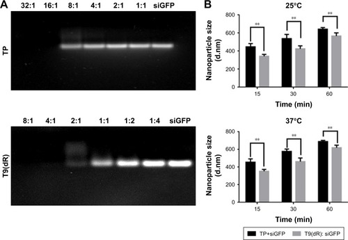
Figure 2 Cell viability treated with complex of T9(dR) or TP and tested by MTT assay. (A) T9(dR) and (B) TP.
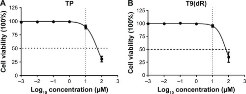
Figure 3 T9(dR) or TP transported siRNA into (A) 293T and (B) A549 cell lines. After T9(dR) or TP and cy3-conjugated siGFP were incubated for 15 minutes, the complex was transfected into cells seeded in eight-well chamber. At 24 hours post-transfection, cells were fixed, mounted with DAPI, and observed under a microscope (20×).
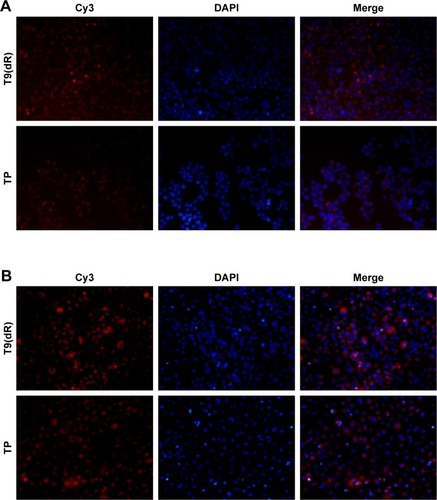
Figure 4 The delivery efficiency of T9(dR) with siRNA in A549, 293T, RAW, and MDCK cells (A–D). After T9(dR) and cy3-conjugated siGFP were incubated for 15 minutes, the complex was transfected into cells seeded in six-well plate. At 24 hours post-transfection, cells were run in a flow cytometer to detect cy3-positive cells. **P-values <0.05 were considered to be significant.
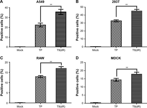
Figure 5 T9(dR) delivered functional siRNA into (A) MDCK and (B) A549 cell lines. MDCK and A549 cells were treated with siNP and infected with influenza virus of MOI =0.01. At 24 hours post-infection, viral titers in supernatant were titrated by standard plaque assay. **P-values <0.05 were considered to be significant.
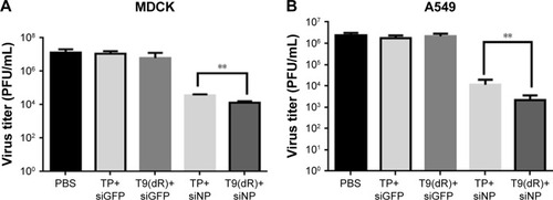
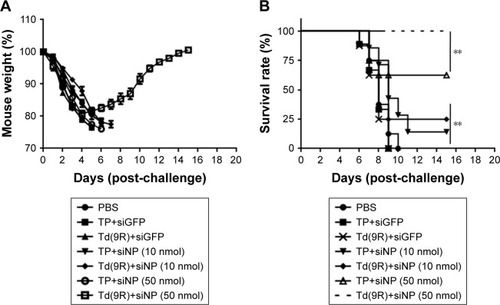
Figure 6 siNP transported by T9(dR) inhibited the replication of PR8 influenza virus in vivo. BALB/c mice were divided into seven groups, intravenously injected PBS, siGFP, siNP with T9(dR) or TP, and infected with influenza. Mouse weight was recorded (A) and survival was calculated (B). **P-values <0.05 were considered to be significant.