Figures & data
Figure 1 Propofol-induced apoptosis in HT-22 cells. (A) CCK-8 assay results showed viability in HT-22 cells treated with 0, 1, 10 and 100 μM propofol, respectively. (B and C) EdU assay results showed EdU-positive HT-22 cells treated with 0, 1, 10 and 100 μM propofol, respectively (B). Quantitative analysis of EdU-positive ratio (C). (D and E) Flow cytometry results showed distribution of apoptotic cells, necrotic cells and survival cells following the treatment of 0, 1, 10 and 100 μM propofol in HT-22 cells, respectively (D). Quantitative analysis of apoptosis rate (E). (F and G) TUNEL results showed TUNEL-positive cells following the treatment of 0, 1, 10 and 100 μM propofol in HT-22 cells, respectively (F). Quantitative analysis of TUNEL-positive rate (G). (H) Protein levels of Bcl-2 and Bax in HT-22 cells treated with 0, 1, 10 and 100 μM propofol, respectively (*p<0.05 compared to control group).
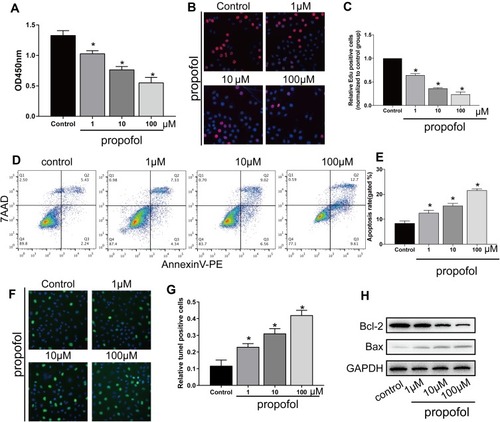
Figure 2 Metformin reversed propofol-induced apoptosis in HT-22 cells (A) CCK-8 assay results showed viability in propofol-induced HT-22 cells either treated with 10 μM metformin or not. (B and C) EdU assay results showed EdU-positive HT-22 cells with propofol induction, followed by 10 μM metformin treatment or not (B). Quantitative analysis of EdU-positive ratio (C). (D and E) Flow cytometry results showed distribution of apoptotic cells, necrotic cells and survival cells in propofol-induced HT-22 cells either treated with 10 μM metformin or not (D). Quantitative analysis of apoptosis rate (E). (F and G) TUNEL results showed TUNEL-positive cells in propofol-induced HT-22 cells either treated with 10 μM metformin or not (F). Quantitative analysis of TUNEL-positive rate (G). (H) Protein levels of Bcl-2 and Bax in propofol-induced HT-22 cells either treated with 10 μM metformin or not (*p<0.05 compared to control group; &p<0.05, compared to propofol (100μM) group).
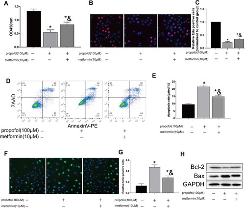
Figure 3 Metformin treatment regulated Cav-1 level. (A and B). Protein level of Cav-1 in HT-22 cells treated with 0, 1, 10 and 100 μM propofol, respectively (A). Grey value analysis of Cav-1 (B). (C and D). Protein level of Cav-1 in propofol-induced HT-22 cells either treated with 10 μM metformin or not (C). Grey value analysis of Cav-1 (D) (*p<0.05 compared to control group; &p<0.05, compared to propofol (100μM) group).
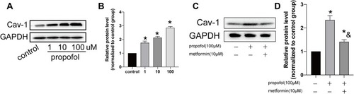
Figure 4 Propofol-induced apoptosis in HT-22 cells through upregulating Cav-1. (A) Transfection efficacy of si-Cav-1 in HT-22 cells. (B) Protein level of Cav-1 in propofol-induced HT-22 cells transfected with si-NC or si-Cav-1. (C) CCK-8 assay results showed viability in propofol-induced HT-22 cells transfected with si-NC or si-Cav-1. (D and E) EdU assay results showed EdU-positive cells in propofol-induced HT-22 cells transfected with si-NC or si-Cav-1 (D). Quantitative analysis of EdU-positive ratio (E). (F and G) Flow cytometry results showed distribution of apoptotic cells, necrotic cells and survival cells in propofol-induced HT-22 cells transfected with si-NC or si-Cav-1 (F). Quantitative analysis of apoptosis rate (G) (*p<0.05 compared to control group; &p<0.05, compared to propofol (100μM) group).
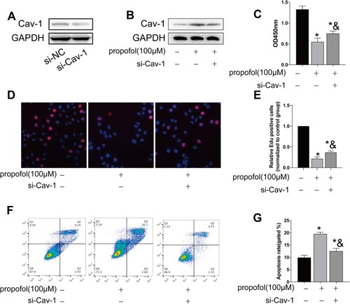
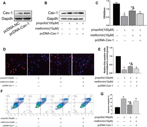
Figure 5 Metformin reversed propofol-induced apoptosis in HT-22 cells through downregulating Cav-1. (A) Transfection efficacy of pcDNA-Cav-1 in HT-22 cells. (B) Protein level of Cav-1 in propofol-induced HT-22 cells transfected with pcDNA-NC or pcDNA-Cav-1 with either metformin treatment or not. (C) CCK-8 assay results showed viability in propofol-induced HT-22 cells transfected with pcDNA-NC or pcDNA-Cav-1 with either metformin treatment or not. (D and E) EdU assay results showed EdU-positive cells in propofol-induced HT-22 cells transfected with pcDNA-NC or pcDNA-Cav-1 with either metformin treatment or not (D). Quantitative analysis of EdU-positive ratio (E). (F and G) Flow cytometry results showed distribution of apoptotic cells, necrotic cells and survival cells in propofol-induced HT-22 cells transfected with pcDNA-NC or pcDNA-Cav-1 with either metformin treatment or not (F). Quantitative analysis of apoptosis rate (G) (*p<0.05 compared to control group; &p<0.05, compared to propofol (100μM) group).