Figures & data
Table 1 The Sequences of ILK siRNA
Figure 1 Emodin inhibited the migration and invasion abilities of endometrial stromal cells. (A). Representative transwell migration and invasion assay of CESs and EESs before and after treated with emodin. (B). Quantification of migration abilities of CESs and EESs before and after treated with emodin (**P < 0.005, ***P < 0.001, ###P < 0.001). (C). Quantification of invasion abilities of CESs and EESs before and after treated with emodin (***P < 0.001, ###P < 0.001).
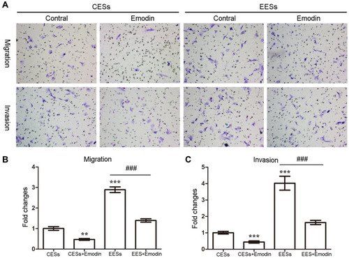
Figure 2 Emodin decreased the expression of ILK, p-GSK-3β and reversed the EMT of endometrial stromal cells in a dose- and time-dependent manner. (A). Representative Western blots showing the expression of ILK, p-GSK-3β, β-catenin, slug, keratin and vimentin in EESs after treated with increasing concentrations of emodin. (B). Quantitative analysis of ILK, p-GSK-3β, β-catenin, slug, keratin and vimentin in EESs after treated with increasing concentrations of emodin (*P < 0.05, **P < 0.005 and ***P < 0.001). (C). Representative Western blots showing the expression of ILK, p-GSK-3β, β-catenin, slug, keratin and vimentin in EESs after treated with emodin for increasing time. (D). Quantitative analysis of ILK, p-GSK-3β, β-catenin, slug, keratin and vimentin in EESs after treated with emodin for increasing time (*P < 0.05, **P < 0.005 and ***P < 0.001).
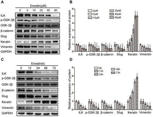
Figure 3 Silencing of ILK inhibited the migration and invasion abilities of EESs by reversing the EMT, which can be strengthened by emodin. (A). Representative Western blots showing the expression of ILK, p-GSK-3β, β-catenin, slug, keratin and vimentin in EESs after transfection siRNA-ILK with or without the treatment with emodin. (B). Quantitative analysis of ILK, p-GSK-3β, β-catenin, slug, keratin and vimentin in EESs after transfection siRNA-ILK with or without the treatment with emodin (*P < 0.05, **P < 0.005, ***P < 0.001, ##P < 0.005, ###P < 0.001). (C). Representative transwell migration and invasion assay of EESs after transfection siRNA-ILK with or without the treatment with emodin. (D). Quantification of migration abilities of EESs after transfection siRNA-ILK with or without emodin (relative to EESs transfected with N.C.; *P < 0.05, **P < 0.005 and ##P < 0.001). (E). Quantification of invasion abilities of EESs after transfection siRNA-ILK with or without emodin (relative to EESs transfected with N.C.; **P < 0.005, ***P < 0.001 and ###P < 0.001).
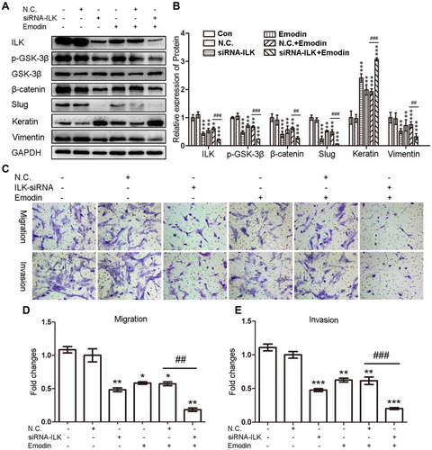
Figure 4 Exogenous expression of ILK facilitated the migration and invasion abilities of CESs by enhancing the EMT, which can be abrogated by emodin. (A). Representative Western blots showing the expression of ILK, p-GSK-3β, β-catenin, slug, keratin and vimentin in CESs after transfection pEGFP-C1-ILK with or without the treatment with emodin. (B). Quantitative analysis of ILK, p-GSK-3β, β-catenin, slug, keratin and vimentin in CESs after transfection pEGFP-C1-ILK with or without the treatment with emodin (relative to CESs transfected with pEGFP-C1; *P < 0.05, **P < 0.005, ***P < 0.001, #P < 0.05,##P < 0.005 and ###P < 0.001). (C). Representative transwell migration and invasion assay of CESs after transfection pEGFP-C1-ILK with or without the treatment with emodin. (D). Quantification of migration abilities of CESs after transfection pEGFP-C1-ILK with or without emodin (relative to CESs transfected with pEGFP-C1; **P < 0.005, ***P < 0.001 and ###P < 0.001). (E). Quantification of invasion abilities of CESs after transfection pEGFP-C1-ILK with or without emodin (relative to CESs transfected with pEGFP-C1; **P < 0.005 and ###P < 0.001).
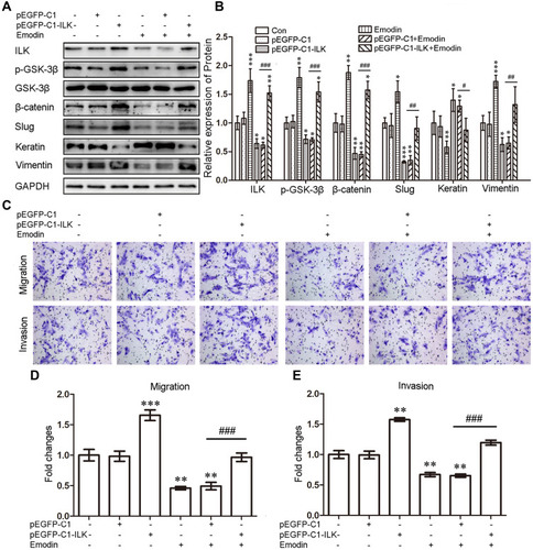
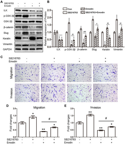
Figure 5 Emodin reversed the EMT of endometrial stromal cells through ILK/GSK-3β pathway. (A). Representative Western blots showing the expression of ILK, p-GSK-3β, β-catenin, slug, keratin and vimentin in CESs after treatment with SB216763 with or without emodin treatment. (B). Quantitative analysis of ILK, p-GSK-3β, β-catenin, slug, keratin and vimentin in CESs after treatment with SB216763 with or without emodin treatment (*P < 0.05, **P < 0.005 and ***P < 0.001, #P < 0.05, ##P < 0.005 and ###P < 0.001). (C). Representative transwell migration and invasion assay of CESs after treatment with SB216763 with or without emodin treatment. (D). Quantification of migration abilities of CESs after treated with SB216763 with or without emodin (*P < 0.05, **P < 0.005 and #P < 0.05). (E). Quantification of invasion abilities of CESs after treated with SB216763 with or without emodin (*P < 0.05, **P < 0.005 and #P < 0.05).