Figures & data
Figure 1 The chemical structure of andrographolide.
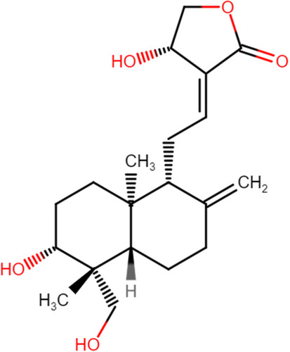
Figure 2 The framework for the selection of relevant studies.
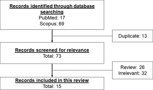
Table 1 In vivo Studies on the Effects of Andrographolide on Myocardial Injuries
Table 2 In vitro Studies on the Effects of Andrographolide on Cardiomyocytes
Figure 3 The mechanism of action of in the regulation of inflammatory response. During myocardial injuries, the overwhelming inflammatory cytokines activate several essential signalling pathways including TLR, NF-κB, PI3K/Akt, MAPK, JAK-STAT signal transduction pathways. The reductions of inflammation by A. paniculata (Burm.f.) Nees and andrographolide are mainly mediated through the inhibition of these signalling pathways (indicated by green arrows). The arrow pointing upward indicates an increase or activation while the arrow pointing downward indicates a decrease or inhibition.
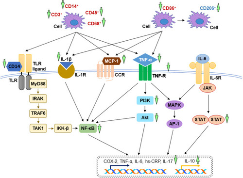
Figure 4 The mechanism of action in the regulation of oxidative stress. During myocardial injuries, the imbalance between ROS/RNS levels and anti-oxidative capacity cause lipid peroxidation and protein modification. The anti-oxidative property A. paniculata (Burm.f.) Nees and andrographolide are accomplished by enhancing the antioxidant system via activation of Nrf-2/Keap-1 pathway as well as decreasing the enzymes responsible for oxidative and nitrosative stress (indicated by green arrows). The arrow pointing upward indicates an increase or activation while the arrow pointing downward indicates a decrease or inhibition.
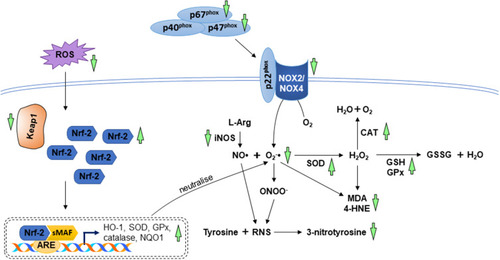
Figure 5 The mechanism of action in the regulation of apoptosis during myocardial injuries. In the extrinsic pathway, the interaction between Fas ligand and its receptor causes the recruitment of FADD and procaspase-8 to form DISC. In the intrinsic pathway, the increases in pro-apoptotic genes and the decrease in anti-apoptotic genes resulted from oxidative stress and cellular damage lead to the release of cytochrome c from mitochondria. Apoptosome formation ensues, consisting of cytochrome c, deoxyadenosine triphosphate (dATP), apoptotic protease-activating factor 1 (Apaf-1), and procaspase-9. The activated initiator caspases (caspase-8 and caspase-9) further activates executioner caspases (caspase-3, caspase-6, and caspase-7), promoting the cleavage of cellular substrate. A. paniculata (Burm.f.) Nees and andrographolide prevent apoptosis by suppressing the mitochondrial and death receptor pathways. The upstream signalling events involved are the activation of IGF-1R and inhibition of PPAR-α. The anti-apoptotic effects of A. paniculata (Burm.f.) Nees and andrographolide are indicated (green arrows). The arrow pointing upward indicates an increase or activation while the arrow pointing downward indicates a decrease or inhibition.
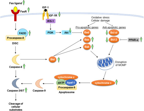
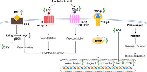
Figure 6 The mechanism of action in the prevention of cardiac fibrosis and endothelial dysfunction. In myocardial injuries, the increased expressions of collagen I, collagen III, fibronectin, PAI-1, and CTGF are mediated through TGF-β/SMAD signalling pathway, leading to the increase in blood coagulation and cardiac fibrosis. High levels of ET-1, TXA2 and low level of PGI2 cause the inhibition of vasodilatation, resulting in endothelial dysfunction. Treatment with A. paniculata (Burm.f.) Nees and andrographolide reverse all these changes, leading to the reduction of cardiac fibrosis, improvement of endothelial function and fibrinolytic function (indicated by green arrows). The arrow pointing upward indicates an increase or activation while the arrow pointing downward indicates a decrease or inhibition.