Figures & data
Figure 1 Vpr expression kinetics and generation of BiFC in transfected cells.
Notes: Cells were cotransfected with equal amounts of VN-Vpr and VC-Vpr plasmids. Hours post-transfection, cells were collected, split into two halves, and used for further analysis. (A) Cells were lysed and analyzed via Western blot using anti-HA antibody to detect Vpr expression. Tubulin was used as loading control. (B) Cells were fixed and analyzed via flow cytometry to detect the percentage of BiFC-positive cells. Results represent one of three independent experiments.
Abbreviations: Vpr, viral protein R; BiFC, bimolecular fluorescence complementation; VN-Vpr, Vpr fused to N-terminus of Venus protein; VC-Vpr, Vpr fused to C-terminus of Venus protein; HA, HA-tagged Vpr.
Abbreviations: Vpr, viral protein R; BiFC, bimolecular fluorescence complementation; VN-Vpr, Vpr fused to N-terminus of Venus protein; VC-Vpr, Vpr fused to C-terminus of Venus protein; HA, HA-tagged Vpr.
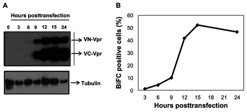
Figure 2 Competition assay to detect the loss of BiFC signal generated by Vpr dimerization.
Notes: Cells were transfected with a constant amount of Venus-Vpr plasmid with increasing concentrations of untagged Vpr plasmid. DNA concentration and volume were normalized using empty vector DNA. Eighteen hours post-transfection, cells were collected and analyzed by flow cytometry. Results represent the mean of four independent experiments, and data from each experiment were normalized to the 1:0 (Venus Vpr: Untagged Vpr) sample.
Abbreviations: BiFC, bimolecular fluorescence complementation; Vpr, viral protein R; DNA, deoxyribonucleic acid.
Abbreviations: BiFC, bimolecular fluorescence complementation; Vpr, viral protein R; DNA, deoxyribonucleic acid.
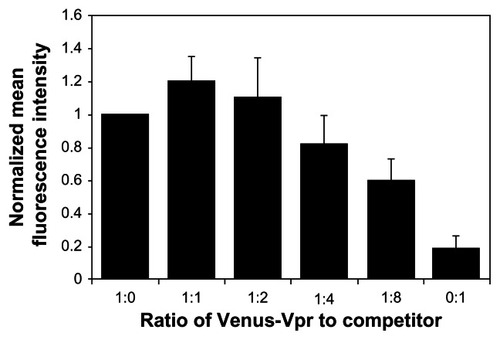
Figure 3 Fluorescence intensity of BiFC generated by Venus-Vpr in the presence of competitor untagged Vpr.
Notes: Cells were seeded onto cover slips and transfected with Venus-Vpr plasmids, Vpr-Flag, or Venus-Vpr and Vpr-Flag. Cells were fixed 18 hours post-transfection, stained with Cy5-conjugated Flag M5 antibody to detect the Vpr competitor molecule. Cells were viewed under confocal microscope at 60X magnification. Blue, DAPI to stain the nucleus; green, BiFC signal to visualize Venus-Vpr; red, Flag-Cy5 signal to detect the competitor Vpr molecule.
Abbreviations: BiFC, bimolecular fluorescence complementation; Vpr, viral protein R; DAPI, 4′,6-diamidino-2-phenylindole, dihydrochloride.
Abbreviations: BiFC, bimolecular fluorescence complementation; Vpr, viral protein R; DAPI, 4′,6-diamidino-2-phenylindole, dihydrochloride.
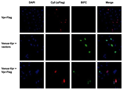
Figure 4 Ability of Vpr peptides to interfere with Vpr dimerization.
Note: (A) Vpr peptides were screened for an effect on nuclear BiFC fluorescence using high content imaging. Cells transfected with Venus-Vpr plasmids were treated with Vpr peptides or vehicle controls (DDDT or PBS) and assessed for BiFC signal as described in methods. Data were normalized to the solvent-appropriate control cells. The average and standard deviation across four independent experiments were calculated and plotted. (B) The average number of viable cells per field, after excluding cells for morphology and nuclear size using the image software, were recorded for each well (based on the average of >20 fields per well). The figure represents one of four independent experiments.
Abbreviations: Vpr, viral protein R; BiFC, bimolecular fluorescence complementation; PBS, phosphate buffered saline; DDDT, dimethyl sulfoxide; TF, transfection.
Abbreviations: Vpr, viral protein R; BiFC, bimolecular fluorescence complementation; PBS, phosphate buffered saline; DDDT, dimethyl sulfoxide; TF, transfection.
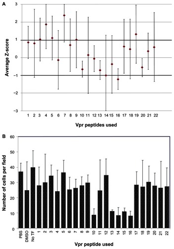
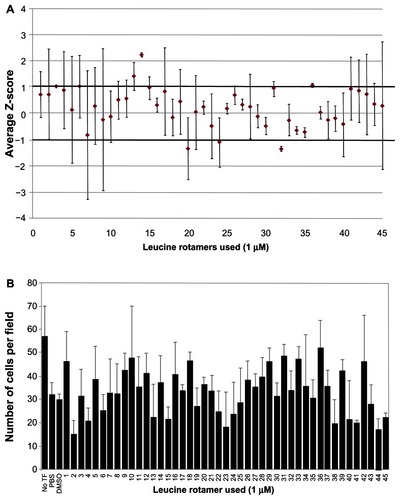
Figure 5 Ability of leucine rotamer library to inhibit Vpr dimerization measured by BiFC signal.
Note: (A) Small molecules (leucine rotamers) were screened for their ability to interfere with Vpr dimerization measured by BiFC fluorescence using high content imaging. Cells transfected with Venus-Vpr plasmids were treated with leucine rotamer library (1 μM) or vehicle control (DDDT) and assessed for BiFC signal. Data was normalized to vehicle exposed transfected cells using Z-score methods. The average and standard deviation from two independent experiments were calculated and plotted. (B) The average number of viable cells per field, after excluding cells for morphology and nuclear size, were recorded for each well (based on the average of >20 fields per well) using the image software. Figure represents one of two independent experiments.
Abbreviations: Vpr, viral protein R; BiFC, bimolecular fluorescence complementation; TF, transfection; PBS, phosphate buffered saline; DDDT, dimethyl sulfoxide.
Abbreviations: Vpr, viral protein R; BiFC, bimolecular fluorescence complementation; TF, transfection; PBS, phosphate buffered saline; DDDT, dimethyl sulfoxide.