Figures & data
Figure 1 A simplified diagram illustrating the structural alterations in human skin affected by psoriasis. Normal skin structure (left) and compared with psoriatic skin (right). The formation of scaly, raised, red plaques on the skin, accompanied by I. acanthosis; II. parakeratosis; III. hyperkeratosis; IV. Munro’s microabscess. These plaques can be uncomfortable and itchy, causing pain and irritation.
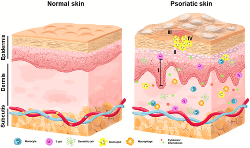
Table 1 List of Non-Coding RNAs in the Pathogenesis of Psoriasis
Figure 2 Gene silencing mechanisms of miRNA. The transcription of miRNA genes occurs in the nucleus via RNA polymerase II, yielding a primary miRNA (pri-miRNA) that is then cleaved by Drosha into a precursor miRNA (pre-miRNA). Exportin 5 mediates the transport of pre-miRNA to the cytoplasm where Dicer cleaves it into mature miRNA. The miRNA is then loaded onto the RISC complex, where the passenger strand is eliminated, and the guide strand directs RISC to partially complementary target mRNA. The binding between the guide strand and target mRNA leads to various modes of target inhibition, including translational repression, degradation, or cleavage.
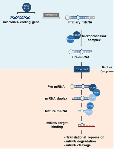
Figure 3 Gene silencing mechanisms of siRNA. Dicer processes dsRNA (either endogenous or exogenous) into siRNA, which is incorporated into the RISC complex. The passenger strand of siRNA is cleaved by AGO2, a component of RISC, leaving the guide strand to direct the complex to the complementary mRNA target. Upon binding, the guide strand initiates cleavage of the target mRNA, leading to gene silencing.
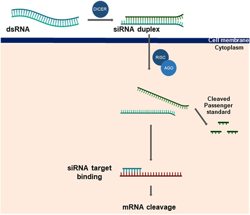
Table 2 Compilation of Previous Studies Investigating miRNA Therapeutics Discovery in Animal Models of Psoriasis
Table 3 Compilation of Previous Studies Investigating siRNA Therapeutics Discovery in Animal Models of Psoriasis
Table 4 List of Other Applications Discovery in Animal Models of Psoriasis
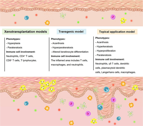
Figure 4 A summary of various methods used to create animal models for studying psoriasis. Various techniques have been utilized to generate animal models of psoriasis, with each method resulting in chronic inflammatory hyperproliferative skin phenotypes that bear resemblance to psoriasis. These manipulations, which involve different cell types and molecular mechanisms, may induce the development of hyperproliferative inflammatory skin changes, suggesting diverse pathogenic mechanisms (elaborated in the text).