76
Views
4
CrossRef citations to date
0
Altmetric
Original Research

Endogenous n-3 polyunsaturated fatty acids (PUFAs) mitigate ovariectomy-induced bone loss by attenuating bone marrow adipogenesis in FAT1 transgenic mice

, , , , , , , , , , , & show all
Pages 545-552 | Published online: 28 Jun 2013

Figures & data

Figure 1 (A and B) Genotyping. (A) PCR genotyping confirmation using FAT1 fragment-specific primers indicated FAT1 gene expression (lane 1) and wild-type (WT) gene expression (lane 2); lane 3 is the negative control. (B) N-6/n-3 ratio of FAT1 mice (mean = 7.762) was significantly lower than WT mice (mean = 28.43).

Note: *P < 0.05 versus FAT1.
Figure 1 (A and B) Genotyping. (A) PCR genotyping confirmation using FAT1 fragment-specific primers indicated FAT1 gene expression (lane 1) and wild-type (WT) gene expression (lane 2); lane 3 is the negative control. (B) N-6/n-3 ratio of FAT1 mice (mean = 7.762) was significantly lower than WT mice (mean = 28.43).

Figure 2 (AE) Gross examination and tissue weight. Three months after ovariectomy, there was increased body weight (C) and abdominal adipose tissue (D) in the ovariectomized (OVX) group compared to the sham group. However, the FAT1 OVX group presented significant lower body weight (A and C) and abdominal adipose tissue (B and D) compared to the wild-type (WT) OVX group. The weight of uterus was significantly lower in the OVX group than the sham group (E).

Notes: *P < 0.05 versus WT OVX;
P < 0.05 versus FAT1 OVX; #
P < 0.05 versus FAT1 OVX.
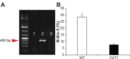
Figure 2 (A–E) Gross examination and tissue weight. Three months after ovariectomy, there was increased body weight (C) and abdominal adipose tissue (D) in the ovariectomized (OVX) group compared to the sham group. However, the FAT1 OVX group presented significant lower body weight (A and C) and abdominal adipose tissue (B and D) compared to the wild-type (WT) OVX group. The weight of uterus was significantly lower in the OVX group than the sham group (E).

Figure 3 (AE) Bone parameter assessment by micro-computed tomography scan. (A) Three-dimensional reconstructions of the distal femoral metaphysis of FAT1 and wild-type (WT) mice by micro-CT scan. The FAT1 ovariectomized (OVX) group exhibited more bone mass than the WT OVX group. (BD) Quantitation of bone parameters of distal femur bone. The values of bone mineral density (BMD, B), bone mineral content (BMC, C), bone volume/total volume (BV/TV, D), and trabecular number (Tb.N) (E) in the OVX group were decreased compared with those in the sham group. The bone parameter (BMD, BMC, BV/TV) values of the FAT1 OVX group were significantly higher than the WT OVX group, but the Tb.N in the FAT1 OVX group only mildly increased than WT OVX group.

Notes: (BE) #
P < 0.05 versus FAT1 OVX; (BE) *P < 0.05 versus WT OVX; (B–E)
P < 0.05 versus FAT1 OVX.
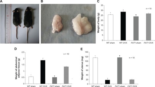
Figure 3 (A–E) Bone parameter assessment by micro-computed tomography scan. (A) Three-dimensional reconstructions of the distal femoral metaphysis of FAT1 and wild-type (WT) mice by micro-CT scan. The FAT1 ovariectomized (OVX) group exhibited more bone mass than the WT OVX group. (B–D) Quantitation of bone parameters of distal femur bone. The values of bone mineral density (BMD, B), bone mineral content (BMC, C), bone volume/total volume (BV/TV, D), and trabecular number (Tb.N) (E) in the OVX group were decreased compared with those in the sham group. The bone parameter (BMD, BMC, BV/TV) values of the FAT1 OVX group were significantly higher than the WT OVX group, but the Tb.N in the FAT1 OVX group only mildly increased than WT OVX group.

Figure 4 (AC) Histological analysis of bone marrow adiposity. (A) Hematoxylin and eosin and toluidine blue staining for distal femur bone marrow. Images were taken at 200x magnification. There were fewer adipocytes in the FAT1 OVX group than the wild-type (WT) ovariectomized (OVX) group. (B) #, per mm2) and (C) adipocyte volume/tissue volume (AV/TV) were significantly reduced in the FAT1 OVX group. Three months after ovariectomy, the OVX group showed more bone marrow adiposity than the sham group (P < 0.05).

Notes: #P < 0.05 versus FAT1 OVX; *P < 0.05 versus WT OVX;
P < 0.05 versus FAT1 OVX. Scale bars, 100 μm
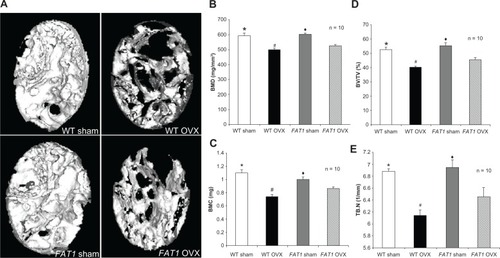
Figure 4 (A–C) Histological analysis of bone marrow adiposity. (A) Hematoxylin and eosin and toluidine blue staining for distal femur bone marrow. Images were taken at 200x magnification. There were fewer adipocytes in the FAT1 OVX group than the wild-type (WT) ovariectomized (OVX) group. (B) #, per mm2) and (C) adipocyte volume/tissue volume (AV/TV) were significantly reduced in the FAT1 OVX group. Three months after ovariectomy, the OVX group showed more bone marrow adiposity than the sham group (P < 0.05).

Figure 5 (AC) Immunohistochemistry and counting of positively stained cells in bone marrow three months after ovariectomy. The black arrows indicate the runt-related transcription factor 2 (RUNX2) positively stained cells, and the red arrows indicate peroxisome proliferator-activated receptor gamma (PPARγ) positively stained cells (200× magnification, scale bar = 100 μm) (A and C). The proportion of RUNX2-positive cells was significantly increased in the FAT1 ovariectomized (OVX) group compared to wild-type (WT mice). (B and C) The proportion of PPARγ-positive cells was significantly reduced in the FAT1 OVX group compared to WT mice.

Notes: #P < 0.05 versus FAT1 OVX; *P > 0.05 versus FAT1 sham; P < 0.05 versus FAT1 OVX;
P > 0.05 versus FAT1 sham. Scale bars, 100 μm
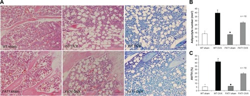
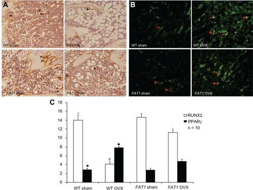
Figure 5 (A–C) Immunohistochemistry and counting of positively stained cells in bone marrow three months after ovariectomy. The black arrows indicate the runt-related transcription factor 2 (RUNX2) positively stained cells, and the red arrows indicate peroxisome proliferator-activated receptor gamma (PPARγ) positively stained cells (200× magnification, scale bar = 100 μm) (A and C). The proportion of RUNX2-positive cells was significantly increased in the FAT1 ovariectomized (OVX) group compared to wild-type (WT mice). (B and C) The proportion of PPARγ-positive cells was significantly reduced in the FAT1 OVX group compared to WT mice.