Figures & data
Table 1 Patient demographics and baseline characteristics
Table 2 Mean BCVA (logMAR)
Table 3 PED height (μm)
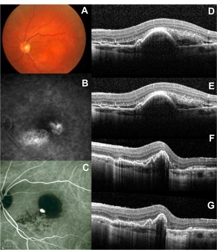
Figure 1 Selected case: A 76-year-old man was referred for decreased visual acuity in the left eye. Baseline examination, including (A) fundoscopy, (B) fluorescein angiography, (C) ICG angiography, and (D) macular OCT revealed a vascular PED associated with exudative AMD. The baseline BCVA and PED height were 20/80 and 380 μm, respectively. The same eye (E) at 3 months (BCVA: 20/70; PED height: 236 μm), (F) at 6 months (BCVA: 20/70; PED height: 176 μm), and (G) at 12 months (BCVA: 20/60; PED height: 166 μm), after the first ranibizumab injection.
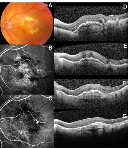
Figure 2 Selected case: A 72-year-old man was referred for decreased visual acuity in the left eye. Baseline examination, including (A) fundoscopy, (B) fluorescein angiography, (C) ICG angiography, and (D) macular OCT revealed a serous PED associated with exudative AMD. The baseline BCVA and PED height were 20/100 and 385 μm respectively. The same eye (E) at 3 months (BCVA: 20/80; PED height: 358 μm), (F) at 6 months (BCVA: 20/70; PED height: 297 μm), and (G) at 12 months (BCVA: 20/60; PED height: 251 μm) after the first ranibizumab injection.