Figures & data
Figure 1 Image after second surgery. Head CT axial image shows the drainage tube placed in the lesion.
Abbreviation: CT, computed tomography.
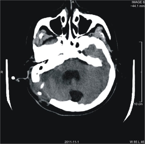
Table 1 Literature on valproic acid (VPA)-associated hypofibrinogenemia
Figure 1 Image after second surgery. Head CT axial image shows the drainage tube placed in the lesion.
Table 1 Literature on valproic acid (VPA)-associated hypofibrinogenemia