Figures & data
Figure 1 The miRNA biogenesis pathway.
Notes: The first step of microRNA biogenesis is the transcription of a long pri-miRNA that contains loop–stem structures. These loop–stem structures will be further cleaved-off, mediated by the Drosha-DGCR8 complex, a processing step that also takes place in the nucleus. The cleavage product is called pre-microRNA and has a typical two nucleotide 3′ overhang, essential for nuclear export by exportin-5. Pre-miRNA constitutes a transport complex together with exportin-5 and its cofactor Ran (the GTP-bound form). Following export, the pre-miRNA is cleaved by Dicer in combination with the TRBP. This yields an approximately 20 base pair miRNA duplex, which is separated and usually one strand is selected as the mature miRNA, whereas the other strand is degraded. After incorporation into miRISC and strand selection, the mature miRNA strand induces translational repression and/or mRNA cleavage, leading to reduction of the protein.Citation18–Citation20
Abbreviations: Ago, argonaute; GTP, guanosine triphosphate; miRISC, miRNA-induced silencing complex; miRNA, microRNA; pri-miRNA, primary miRNA; pre-miRNA, precursor miRNA; TRBP, trans-activation response element RNA binding protein.
Abbreviations: Ago, argonaute; GTP, guanosine triphosphate; miRISC, miRNA-induced silencing complex; miRNA, microRNA; pri-miRNA, primary miRNA; pre-miRNA, precursor miRNA; TRBP, trans-activation response element RNA binding protein.
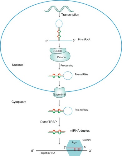
Figure 2 Putative mechanisms of α-synuclein neurodegeneration in familial PD.
Notes: The presence of Lewy bodies in PD is caused by a failure to properly dispose of α-synuclein. Point mutations in α-synuclein and the recently described Iowan functional duplication lead to excessive intracellular accumulation of α-synuclein, resulting in spontaneous oligomerization of this protein. Mutant α-synuclein can inhibit proteasomal activity, potentially leading to enhanced accumulation; soluble α-synuclein protofibrils and intracellular protein aggregates have been shown to impair the proteasome. Dopamine may also form stable adducts with protofibrils, potentially enhancing this toxicity. Genetic mutations associated with the UPP pathway, including parkin and UCH-L1, are associated with familiar PD. Combined with other cellular stress, this leads to neuronal loss.
Abbreviations: PD, Parkinson’s disease; UCH-L1, ubiquitin carboxy-terminal hydrolase L1; UPP, ubiquitin proteasome pathway.
Abbreviations: PD, Parkinson’s disease; UCH-L1, ubiquitin carboxy-terminal hydrolase L1; UPP, ubiquitin proteasome pathway.
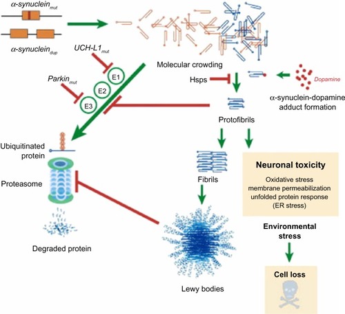
Figure 3 MicroRNA (miR-7 and miR-153) regulation of SNCA in PD.
Notes: The brain-enriched miRNAs, miR-7 and miR-153, are predicted to bind to the α-syn mRNA 3′-UTR as their targets. There is a significant synergy effect between miR-7 and miR-153 in down regulating α-syn mRNA levels and protein expressions. On the contrary, depletion of these miRNAs results in a concomitant increase in α-syn levels in a PD brain. miR-7 has a protective role by preventing oxidative stress and miR-7 inhibition causes cell death.
Abbreviations: 3′-UTR, 3′ untranslated region; α-syn, α-synuclein; mRNA, messenger RNA; miRNA, microRNA; PD, Parkinson’s disease.
Abbreviations: 3′-UTR, 3′ untranslated region; α-syn, α-synuclein; mRNA, messenger RNA; miRNA, microRNA; PD, Parkinson’s disease.

Figure 4 LRRK2 regulates the microRNA (let-7 and miR-184*) network.
Notes: Pathogenic LRRK2 inhibited let-7 and miR-184* function and consequently transcription factors E2F1 and DP were upregulated, and such effects were dependent on LRRK2 kinase activity. Overexpression of E2F1 and DP may lead to dopamine neuron degeneration and impaired locomotor activity.
Abbreviation: LRRK2, leucine-rich repeat kinase 2.
Abbreviation: LRRK2, leucine-rich repeat kinase 2.
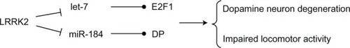
Figure 5 The role of PARK2 and PARK7 in the pathogenesis of PD.
Notes: In accordance with numerous studies, the genes PARK2 and PARK7 are also involved in the pathogenesis of PD. PARK2 encodes the parkin protein, and the DJ-1 protein is encoded by PARK7. In the brains of PD patients, the levels of miR-34b and miR-34c are significantly decreased, decreased levels of miR-34b and miR-34c are also accompanied by decreased concentrations of parkin and DJ-1 proteins. Nevertheless, there is no experimental evidence for miR-34b/miR-34c targeting to genes encoding parkin and DJ-1.
Abbreviation: PD, Parkinson’s disease.
Abbreviation: PD, Parkinson’s disease.
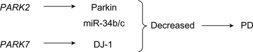
Figure 6 miR-133b and Pitx3 form a negative feedback loop.
Notes: Summary of the regulation of DA neuronal events in the brain within a negative feedback loop. The transcription factor Pitx3 induces transcription of miR-133b during neural differentiation, which in turn represses Pitx3.
Abbreviations: DA, dopaminergic; Pitx3, pituitary homeobox 3.
Abbreviations: DA, dopaminergic; Pitx3, pituitary homeobox 3.
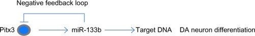
Figure 7 MicroRNA (miR-433) regulation of FGF20 in PD.
Notes: FGF20 disruptions are associated with a higher risk of PD, and one of its SNPs is within a miR-433 binding site. Decreased miR-433 binding efficiency results in FGF20 overexpression and a concomitant increase in α-syn protein levels.
Abbreviations: α-syn, α-synuclein; FGF20, fibroblast growth factor 20; PD, Parkinson’s disease; SNP, single-nucleotide polymorphism.
Abbreviations: α-syn, α-synuclein; FGF20, fibroblast growth factor 20; PD, Parkinson’s disease; SNP, single-nucleotide polymorphism.
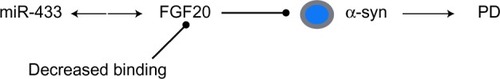
Table 1 miRNAs potentially involved in PD pathophysiology
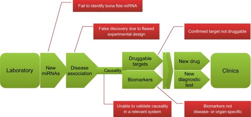
Figure 8 The road from laboratory to clinic: the promises and challenges of miRNA research.
Notes: The hopscotch course in green is a layout of an ideal path of miRNA research evolved from basic research to clinical practice. Red boxes indicate major challenges at different steps.
Abbreviation: miRNA, microRNA.
Abbreviation: miRNA, microRNA.