Figures & data
Figure 1 Dried particles adhere to the drying chamber.
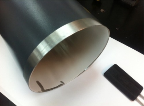
Figure 2 Narrow distribution of particle size for the gelatin vildagliptin nanoparticles formulation prepared by the spray-drying technique.
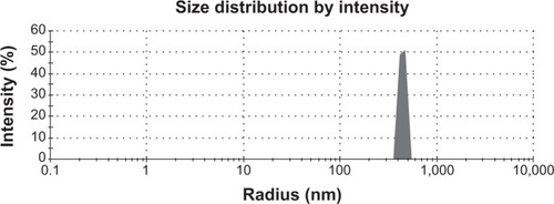
Figure 3 Surface morphology of the gelatin vildagliptin nanoparticles preparation showing a smooth surface and surface-folding shape.
Figure 4 Percentage swelling profile.
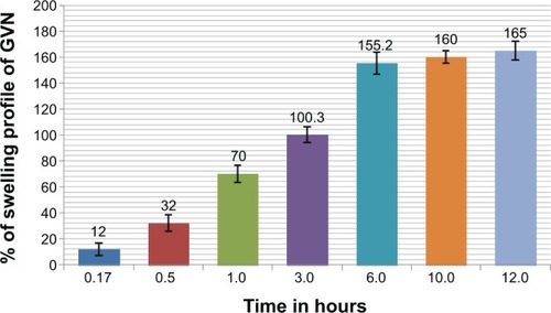
Figure 5 In vitro release testing of vildagliptin and the gelatin vildagliptin nanoparticles (GVN) preparation – Peppas model.
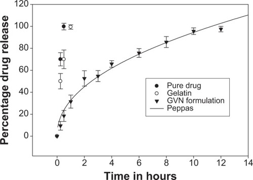
Figure 6 Fourier-transform infrared spectra of vildagliptin, aminated gelatin, and nanoparticle formulation.
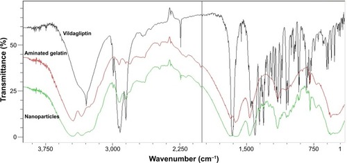
Figure 7 The percentage of gelatin vildagliptin nanoparticles (GVN) retained in the gastrointestinal tract for longer periods of time compared to active pharmaceutical ingredients.
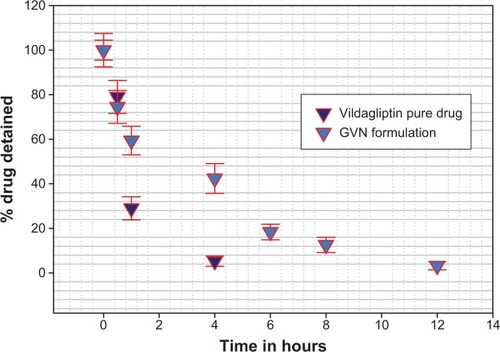
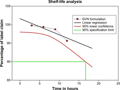
Figure 8 Vildagliptin-content plot for the gelatin vildagliptin nanoparticles (GVN) formulation (real-time storage conditions).