
Figures & data
Figure 1 Characteristics and targets of frequently used species in animal models of pneumonia.
Notes: The main animal species used are presented. For each, the experimental advantages are mentioned in a panel followed by the list of the main topic for which these species are usually used.
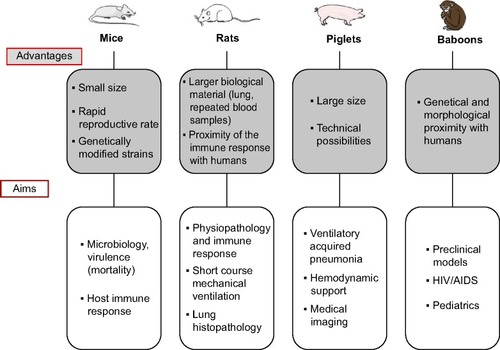
Figure 2 Whole-body inhalation exposure system.
Notes: The Glas-Col® aerosol exposure chamber system (Glas-Col, Terre Haute, IN, USA) is presented here. The bacterial suspension at a known concentration (A) is placed into a glass vial that is a venturi nebulizer (B), connected to a leak-proof gas flow system (C). Pumps generate compressed gas flow to disperse the suspension into a fine mist. Pumps’ flow rate is adjusted on manual flowmeters (D). Durations of the aerosolization and decontamination time are set via digital interface software displayed on a screen window (E). High-efficiency particulate arrestance filters, gas incinerators, and UV decontamination (F) processes are all included in a whole block. The system operates under negative pressure. Before aerosolization starts, animals are placed in a stainless steel basket (G).
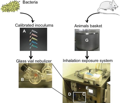
Figure 3 Chronic pneumonia model using agar beads: main steps.
Notes: (A) Agar beads synthesis (top row of pictures). Broth containing bacteria and agar is added to mineral oil with continuous shaking and heating. The solution obtained is centrifuged to obtain beads whose size is measured and must be about 100 µM. The precise inoculum is assessed by serial dilutions method. (B) Model of rat pneumonia (bottom row of pictures). After inhaled anesthesia, the rat is suspended by the teeth and intubated into the trachea. Agar beads solution is injected into a tracheal catheter. After animal sacrifice, macroscopic aspects of lung lesions can be observed. Then, the lungs are homogenized for bacterial count.
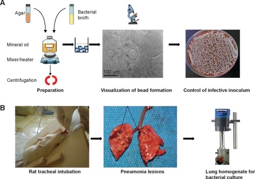
Figure 4 Histological pictures.
Notes: Magnification ×30 showing lung lesions of acute (A) and chronic Pseudomonas aeruginosa pneumonia. Chronic pneumonia was obtained with infected agar beads intra-tracheal inoculation (B). Black arrows indicate the lesions of bronchopneumonia. (C) Shows normal lung and (D) is issued from rat lungs inoculated with sterile agar beads and shows normal lung parenchyma.
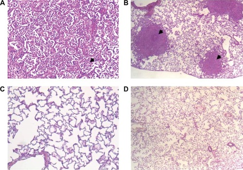
Figure 5 Pathophysiological mechanisms explaining the severity of a Streptococcus pneumoniae pneumonia following an influenza virus lung infection according to mice models.
Notes: When influenza pneumonia is followed by a streptococcal superinfection, the severity resulting from the synergy of both microorganisms can be observed at different levels. In the lungs, it is associated with edema, inflammation, and impaired immune response. From a microbiological point of view, the bacterial clearance is decreased and lung abscesses are more frequently described. extra-pulmonary bacterial dissemination is also more favored with an increased occurrence of bacteremia.
Abbreviation: A1AT, alpha1 antitrypsine; LYZ, lysozyme; NE, neutrophil elastase; LT, T lymphocytes.
Abbreviation: A1AT, alpha1 antitrypsine; LYZ, lysozyme; NE, neutrophil elastase; LT, T lymphocytes.
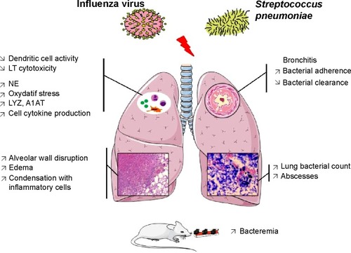
Table 1 Characteristics of the major pathogen associations described in animal models of pneumonia