Figures & data
Figure 1 Alteration in numbers and phenotype of DCs in TDLNs after cryo-ablation in glioma mice models.
Notes: (A) Scheme to study the alteration of DCs in glioma mice models. GL261 cells (106) were implanted intradermally in the left flank 14 days prior to day 0. Two weeks later, when tumors grew to 5 to 9 mm, mice were randomized into tumor bearing group, cryo-ablation group, and surgery group. After 14 days, TDLNs were removed and DCs were isolated. The second tumors were implanted on day 28. (B, C) Expression levels of CD80, CD86, and MHC class II in gated CD11c+ DCs from four groups were evaluated by FACS analysis. Grey curves represent staining with the isotype-matched control monoclonal antibodies. (D) Absolute amounts of DCs were calculated. The data are representative of three independent experiments. Statistical significance was calculated by one-way ANOVA using the Bonferroni post-test with the following notations for P-value. *P<0.05, error bars, SEM. #Day –14 is 14 days prior to day 0.
Abbreviations: DCs, dendritic cells; TDLNs, tumor-draining lymph nodes; DLN, draining lymph node; FACS, fluorescence activated cell sorting; ANOVA, analysis of variance; SEM, standard error of the mean; MHC, major histocompatibility complex; FITC, fluorescein isothiocyanate.
Abbreviations: DCs, dendritic cells; TDLNs, tumor-draining lymph nodes; DLN, draining lymph node; FACS, fluorescence activated cell sorting; ANOVA, analysis of variance; SEM, standard error of the mean; MHC, major histocompatibility complex; FITC, fluorescein isothiocyanate.
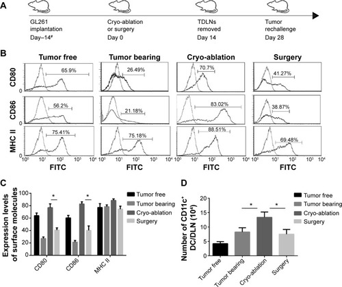
Figure 2 Cryo-ablation converted the expression of IL-10 in TDLN-DCs.
Notes: (A) Expressions of intracellular IL-10 in gated CD11c+ DCs from four groups were evaluated by FACS analysis. The upper left quadrant represents the IL-10+ DCs, upper right quadrant represents the IL-10+CD11c+ DCs, lower right quadrant represents the CD11c+ DCs, the lower left quadrant represents the IL-10− CD11c− DCs. (B) Percentages of IL-10+CD11c+ were calculated in the histogram. *P<0.05 represents the IL-10 levels in the cryo-ablation group compared to tumor bearing or surgery group in (C). (C) Ex vivo IL-10 secretion by 104 DCs in overnight culture, determined by ELISA, presented as the mean plus SEM of triplicate wells. The data were analyzed by Student’s t-test. *P<0.05 represents the IL-10+ DC cells in the cryo-ablation group compared to tumor bearing or surgery group in .
Abbreviations: IL, interleukin; TDLN, tumor-draining lymph node; DCs, dendritic cells; FACS, fluorescence activated cell sorting; ELISA, enzyme-linked immunosorbent assay; SEM, standard error of the mean; MHC, major histocompatibility complex; FSC, forward scatter; SSC, side scatter.
Abbreviations: IL, interleukin; TDLN, tumor-draining lymph node; DCs, dendritic cells; FACS, fluorescence activated cell sorting; ELISA, enzyme-linked immunosorbent assay; SEM, standard error of the mean; MHC, major histocompatibility complex; FSC, forward scatter; SSC, side scatter.
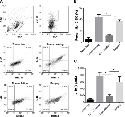
Figure 3 The capacity of DCs to stimulate T cells enhanced after cryo-ablation.
Notes: (A) Mixed lymphocyte responses were performed. Responder CD3+T cells were from BALB/c mice, labeled with CFSE, and incubated with the different DCs. Following incubation, the proliferation levels of cells were analyzed by FACS. (B) T cells were proliferated at various DCs:T cells ratios. (C) T cells stimulated with different DCs from four groups showed cytotoxicity to GL261 cells at various E:T ratios. (D) T cells stimulated with these DCs plus GL261 lysates showed cytotoxicity to GL261 cells at various E:T ratios. The data are representative of three independent experiments. Statistical significance was calculated by one-way ANOVA using the Bonferroni post-test with the following notations for P-value. *P<0.05, error bars, SEM.
Abbreviations: DCs, dendritic cells; CFSE, carboxyfluorescein diacetate succinimidyl ester; FACS, fluorescence activated cell sorting; ANOVA, analysis of variance; SEM, standard error of the mean; FITC, fluorescein isothiocyanate.
Abbreviations: DCs, dendritic cells; CFSE, carboxyfluorescein diacetate succinimidyl ester; FACS, fluorescence activated cell sorting; ANOVA, analysis of variance; SEM, standard error of the mean; FITC, fluorescein isothiocyanate.
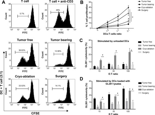
Figure 4 Cryo-ablation could delay tumor growth in tumor rechallenge and reduce the expression of IL-10 in tumor-infiltrating DCs.
Notes: (A) Mice were re-injected 104 GL261 cells in the left flank. Tumors were measured every 4 to 5 days using Vernier calipers. Data are pooled from five mice/group. The tumor size in each group was also analyzed and compared between groups. (B) Second tumor-free survival was plotted as a Kaplan–Meier curve and the log rank test was used for statistical analysis. However, no significant difference was discovered in the tumor-free survival time of these three groups. (C) Immunofluorescence microscopy of 20 μm thick sections of second tumors from surgery group (top) and cryo-ablation group (bottom) were stained for CD11c (red) and IL-10 (green). DAPI was blue. Scale bar: 20 μm. The data are representative of three independent experiments. Statistical significance was calculated by one-way ANOVA using the Bonferroni post-test with the following notations for P-value. *P<0.05, error bars, SEM.
Abbreviations: IL, interleukin; DCs, dendritic cells; DAPI, 4′,6-diamidino-2-phenylindole; ANOVA, analysis of variance; SEM, standard error of the mean.
Abbreviations: IL, interleukin; DCs, dendritic cells; DAPI, 4′,6-diamidino-2-phenylindole; ANOVA, analysis of variance; SEM, standard error of the mean.
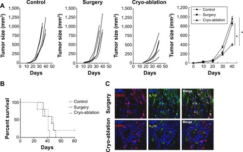
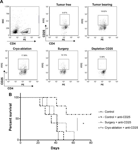
Figure 5 Depletion of Tregs improves the survival rate of cryo-ablated mice.
Notes: (A) Percentages of CD25+CD4+Tregs in CD4+T cells from groups were shown by FACS. (B) Depletion of CD25+CD4+Tregs improves anti-tumor effect in cryo-ablation group. Kaplan–Meier curve showing tumor-free survival of mice with surgery, cryo-ablation, and no treatment with depletion of CD25+CD4+Tregs. Data were pooled from three independent experiments. The log rank test was used for statistical analysis. *P<0.05 represents survival time of the cryo-ablation and anti-CD25 group compared to the other three groups in B.
Abbreviations: FACS, fluorescence activated cell sorting; FITC, fluorescein isothiocyanate; PE, phycoerythrin; SSC, side scatter.
Abbreviations: FACS, fluorescence activated cell sorting; FITC, fluorescein isothiocyanate; PE, phycoerythrin; SSC, side scatter.