Figures & data
Figure 1 A photographic image of the surgical procedure showing the gap between the bone segments after fixing the microplate.
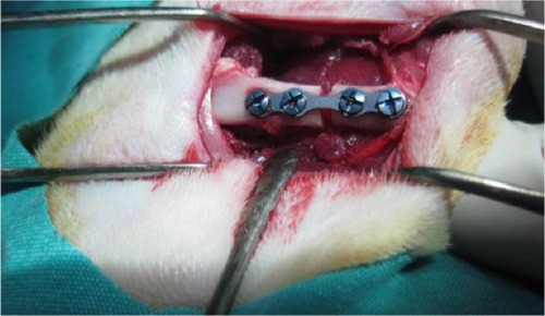
Table 1 Statistical analysis of bone healing at Day 21 and Day 30
Figure 2 Comparison of histopathological bone-healing scores among groups according to the Huo scale.
Notes: *Contradictory values; – median value of control 21 group.
Abbreviations: EMF, electromagnetic field; PTX, pentoxifylline; MIX, both EMF and PTX treatment.
Abbreviations: EMF, electromagnetic field; PTX, pentoxifylline; MIX, both EMF and PTX treatment.
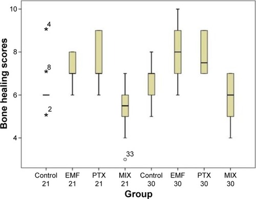
Figure 3 Histopathological sample of bone tissue. The sample shows mostly cartilage and a little newly formed, immature bone tissue.
Notes: The sample is stained with hematoxylin and eosin; magnification, 200×.
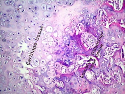
Figure 4 Histopathological sample of bone tissue. The sample shows mostly immature bone tissue.
Notes: The sample is stained with hematoxylin and eosin; magnification, 200×.
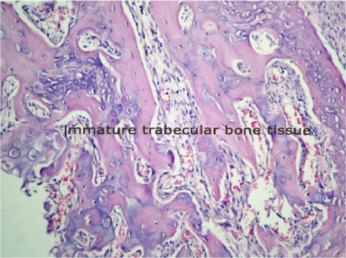
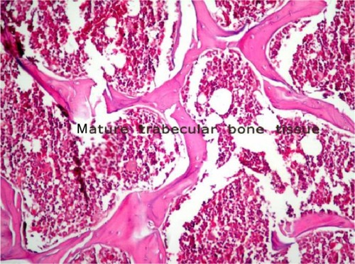
Figure 5 Histopathological sample of bone tissue. The sample is mostly mature bone tissue.
Notes: The sample is stained with hematoxylin and eosin; magnification, 200×.