
Figures & data
Figure 1 Islets transplantation imaging of MRI, SPECT and PET. (A) In vivo MR imaging of recipients having transplanted islets. In vivo spin echo T2-weighted axial MR images of heparin-SPIO-conjugated islets 30 days after xenotransplantation under the renal subcapsular membrane of left kidney in nude mice (300 islet equivalent/mouse). The dark area in the left kidney represents a labeled islet graft. Arrow: transplantation site. Reprinted from Biomaterials. 32(35). Jung MJ, Lee SS, Hwang YH, et al.MRI of transplanted surface-labeled pancreatic islets with heparinized superparamagnetic iron oxide nanoparticles. 9391–9400, Copyright (2011), with permission from Elsevier.Citation21 (B) Transplanted islets under the left kidney capsule of CD1 mice labeled by 111In-tropolone was imaged by three-dimensional reconstructions SPECT/CT. This research was originally published in J Nucl Med. Tai JH, Nguyen B, Wells RG, et al. Imaging of gene expression in live pancreatic islet cell lines using dual-isotope SPECT. J Nucl Med. 2007;49(1):94–102. ©SNMMI. http://jnm.snmjournals.org/content/49/1/94.short.Citation39 (C) PET images were obtained after the [68Ga]DO3A-VS-Cys40-Exendin-4 intravenous injections via the tail in NOD/SCID mice with human-transplanted islets in the liver. The liver with transplanted islets demonstrate prominent tracer uptake (arrow). Reproduced from Junfeng L, Rawson J, et al. Evaluation of [68Ga]DO3A-VSCys40- exendin-4 as a PET probe for imaging human transplanted islets in the liver. Sci Rep. 2019;9:5705. Creative Commons license and disclaimer available from: http://creativecommons.org/licenses/by/4.0/legalcode.Citation56
![Figure 1 Islets transplantation imaging of MRI, SPECT and PET. (A) In vivo MR imaging of recipients having transplanted islets. In vivo spin echo T2-weighted axial MR images of heparin-SPIO-conjugated islets 30 days after xenotransplantation under the renal subcapsular membrane of left kidney in nude mice (300 islet equivalent/mouse). The dark area in the left kidney represents a labeled islet graft. Arrow: transplantation site. Reprinted from Biomaterials. 32(35). Jung MJ, Lee SS, Hwang YH, et al.MRI of transplanted surface-labeled pancreatic islets with heparinized superparamagnetic iron oxide nanoparticles. 9391–9400, Copyright (2011), with permission from Elsevier.Citation21 (B) Transplanted islets under the left kidney capsule of CD1 mice labeled by 111In-tropolone was imaged by three-dimensional reconstructions SPECT/CT. This research was originally published in J Nucl Med. Tai JH, Nguyen B, Wells RG, et al. Imaging of gene expression in live pancreatic islet cell lines using dual-isotope SPECT. J Nucl Med. 2007;49(1):94–102. ©SNMMI. http://jnm.snmjournals.org/content/49/1/94.short.Citation39 (C) PET images were obtained after the [68Ga]DO3A-VS-Cys40-Exendin-4 intravenous injections via the tail in NOD/SCID mice with human-transplanted islets in the liver. The liver with transplanted islets demonstrate prominent tracer uptake (arrow). Reproduced from Junfeng L, Rawson J, et al. Evaluation of [68Ga]DO3A-VSCys40- exendin-4 as a PET probe for imaging human transplanted islets in the liver. Sci Rep. 2019;9:5705. Creative Commons license and disclaimer available from: http://creativecommons.org/licenses/by/4.0/legalcode.Citation56](/cms/asset/617ebe0a-5d95-465d-a513-83b9038097ea/dmso_a_12172308_f0001_c.jpg)
Figure 2 Islets transplantation imaging of FI, BLI and US. (A) Transplanted rat islets were detected by PiF fluorescence imaging. Reprinted with permission from Kang NY, Lee JY, Lee SH, et al. Multimodal imaging probe development for pancreatic β cells: from fluorescence to PET. J Am Chem Soc. 2020;142(7):3430–3439. Copyright (2020) American Chemical Society.Citation79 (B) 500 human islets were transduced with Adeno-CMV-Luc and implanted under the left kidney capsule of NOD-SCID mice. A representative CCD image 3 days postimplantation is shown. Reprinted from Mol Ther. 9(3). Lu Y, Dang H, Middleton B, et al. Bioluminescent monitoring of islet graft survival after transplantation. 428–435, Copyright 2004, with permission from Elsevier.Citation65 (C) Intraoperative ultrasound findings of the portal vein. The transplanted islets appeared as hyperechoic clusters in the portal vein (arrows). Reproduced from Sakata N, Goto M, Gumpei Y, et al. Intraoperative ultrasound examination is useful for monitoring transplanted islets: a case report. Islets. 2012;4(5):339–342, reprinted by permission of the publisher (Taylor & Francis Ltd, hhtp://www.tandfonline.com).Citation70
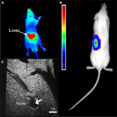
Figure 3 Trimodal imaging of transplanted pancreatic islets in scaffolds. Representative (A) bioluminescence, (B) fluorescence, and (C) axial F-19/H-1 MR images of 3000 and 1000 pancreatic islets transplanted into scaffolds on days 4. Reproduced from Gálisová A, Herynek V, Swider E, et al. A trimodal imaging platform for tracking viable transplanted pancreatic islets in vivo: F-19 MR, fluorescence, and bioluminescence imaging. Mol Imaging Biol. 2019;21(3):454Y464. Creative Commons license and disclaimer available from: http://creativecommons.org/licenses/by/4.0/legalcode.Citation11
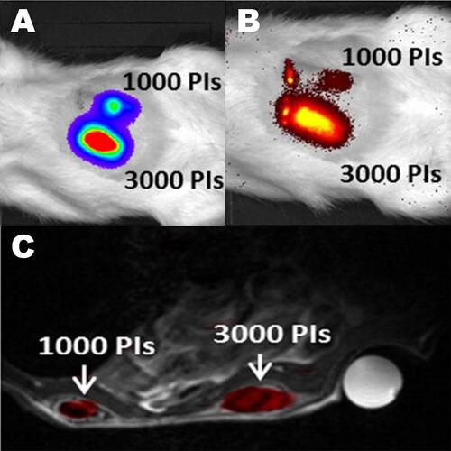
Table 1 Advantages and Disadvantages of Imaging Modalities of Islets