Figures & data
Table 1 Inclusion and Exclusion Criteria
Figure 1 The comparison of one patient in the treatment group before and after treatment. (A) At admission; (B) before enrollment, no obvious necrotic tissue on the wound after debridement; (C) after the second cycle of negative-pressure wound therapy (NPWT); (D) after the fourth cycle of NPWT; (E) the application of the adhesive non-invasive skin stretching device; (F) wound healing.
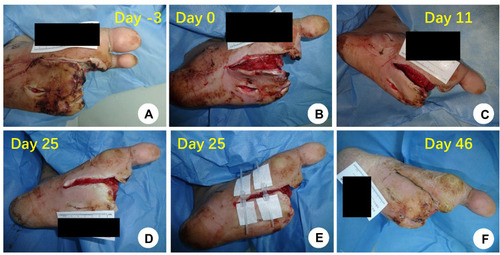
Figure 2 (A–D) A comparison of one patient in the negative-pressure wound therapy (NPWT) + non-invasive skin stretching device (NSSD) group before and after treatment. (A) Before enrollment, no obvious necrotic tissue on the wound after debridement; (B) after NPWT; (C) application of the adhesive NSSD; (D) wound healing. (E–H) A comparison of one patient in the NPWT + Control group before and after treatment; (E) before enrollment, no obvious necrotic tissue on the wound after debridement; (F) during the NPWT; (G) after the NPWT; (F) wound healing.
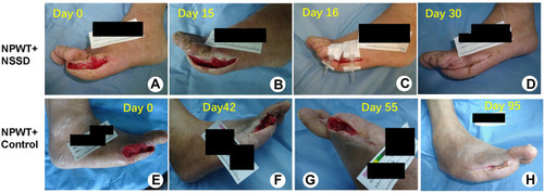
Table 2 Comparison Between Treatment Groups of Patient and Wound Demographics, Comorbidities, and Medications [±s or Median (The Upper and Lower Quartiles) or Case (Composition Ratio)]
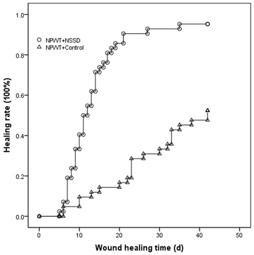
Figure 3 A comparison of the Kaplan–Meier healing curve between the two groups; P < 0.01.
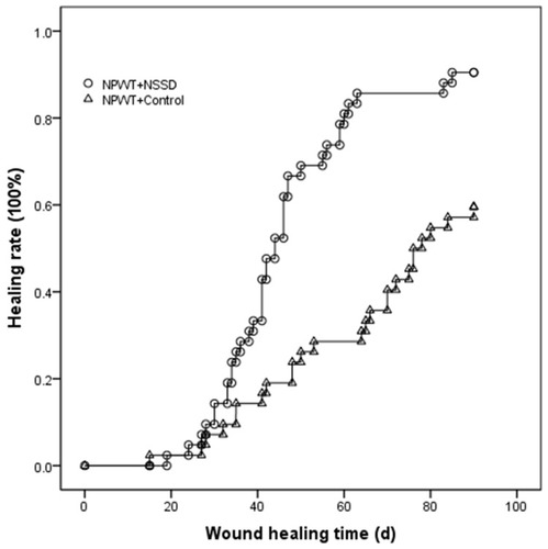
Figure 4 The time-healing Kaplan–Meier curves for both groups with the end of the final negative-pressure closed drainage as the time starting point; P < 0.01.