
Figures & data
Figure 1 Pancreas, intestinal tissues, and microflora in normal and T1DM conditions.
Notes: (A) Under normal conditions, the pancreas is in a sterile environment, the intestinal tissue structure is complete, and the intestinal flora is primarily restricted to the intestinal cavity. (B) In T1DM, intestinal permeability is increased, the mucus layer thickness is reduced, and bacterial contact with intestinal epithelial cells or transfer to other tissues or organs through damaged intestinal epithelial cells occur. 1: Mucosal immune response is induced after bacterial contact with intestinal epithelial cells; 2: Bacterial transfer to the pancreas causes an immune response that can damage the pancreas.
Abbreviations: APC, antigen-presenting cell; Th, helper T cell.
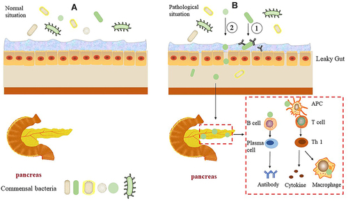
Figure 2 Communication between intestinal microbiota and immune cells.
Notes: SFB can promote the proliferation of Th17 cells in the lamina propria through colonization of the intestinal mucosa. Following their activation, DCs collect B. fragilis and PSA from the intestinal cavity and migrate to lymphatic organs, promoting Th1 differentiation by releasing IL-12. Clostridium colonization in the gut can produce butyrate, which induces Treg expansion. Meanwhile, acetate significantly reduced the number of autoreactive T cells.
Abbreviations: SFB, segmented filamentous bacteria; DC, dendritic cells; IL-12, interleukin 12; Th, helper T cell; Treg, regulatory T cell.
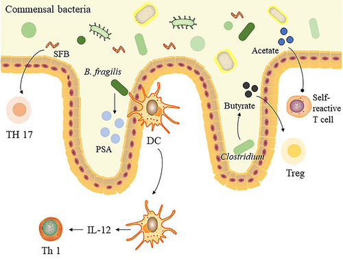
Table 1 The Effect of Treatments on Intestinal Microbiota in T1DM Clinical Trial