Figures & data
Table 1 Comparisons of Clinical Parameters Among NC Group, T2DM Group, and DFU Group [n (%), (±S), M (P25, P75)]
Table 2 Relationship Between miR-155 Expression Levels in Peripheral Blood and Clinical Characteristics of DFU [n (%)]
Table 3 Relationship Between miR-155 Expression Levels in Wound Margin Tissue and Clinical Characteristics of DFU [n (%)]
Figure 1 Curve of wound complete healing rate of DFU between miR-155 high expression group and low expression group by Kaplan-Meier survival curve analysis. (A) The cumulative rate of unhealed DFU in the high expression group of peripheral blood miR-155 was higher than that in the low expression group of peripheral blood miR-155 (log rank, P = 0.004). The estimated time median of wound complete healing in peripheral blood miR-155 high expression group and low expression group were 10.23 weeks and 9.51 weeks, respectively (P < 0.05). (B) The cumulative rate of unhealed DFU in the high expression group of wound margin tissue miR-155 was higher than that in the low expression group of wound margin tissue miR-155 (log rank, P < 0.001). The estimated time median of wound complete healing in wound margin tissue miR-155 high expression group and low expression group were 10.12 weeks and 8.67 weeks, respectively (P < 0.05).
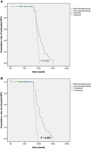
Table 4 Correlations Between miR-155 Expression in Peripheral Blood and Other Clinical Parameters in NC Group, T2DM Group, and DFU Group (r)
Table 5 Correlation Between miR-155 Expression in Wound Margin Tissue and Other Clinical Parameters in the DFU Group (r)
Table 6 The Multivariate Stepwise Logistic Regression Analysis of Risk Factors for Diabetic Foot Ulcer
Figure 2 The biomarker potential of circulating miR-155 for DFU and control. ROC analysis was used to evaluate the ability of circulating miR-155 to distinguish between two groups. MiR-155 distinguished DFU patients from controls with area under curve (AUC) of 0.794 (95% CI, 0.726–0.863, P < 0.001), the best cut-off point of miR-155 was 1.01, the sensitivity was 96.82%, and the specificity was 95.93%. DFU: diabetic foot ulcer.
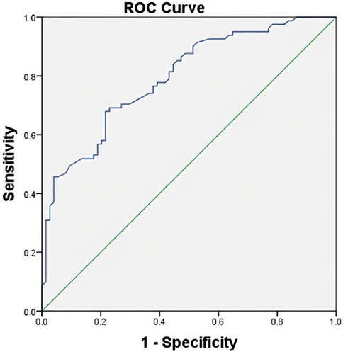
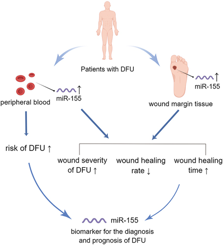
Figure 3 MiR-155 is a potentially valuable biomarker for diagnosis and prognosis of DFU. ↑: increase; ↓: decrease.