Figures & data
Figure 1 Schematic illustration of the experimental procedures.
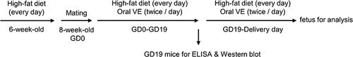
Figure 2 Vitamin E (VE) ameliorates diabetes-induced glucose and insulin intolerance in pregnant mice. (A) Blood glucose levels in pregnant wild-type (WT) and gestational diabetes mellitus (GDM) mice treated water and VE in different concentrations (n=6 mice). (B) The area under the curve for blood glucose levels during the oral glucose tolerance test (OGTT) (n=6 mice). (C) Serum insulin levels in pregnant WT and GDM mice from 0 to 30 minutes following OGTT (n = 6 mice). Values are expressed as means ± SD. *p < 0.05, ****p < 0.0001.
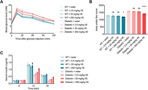
Figure 3 Vitamin E (VE) inhibits dysregulation of adipocytokine expression and hyperlipidemia in pregnant gestational diabetes mellitus (GDM) mice. (A) Immunoblot analysis of adiponectin, interleukin (IL)-6, the cell-bound precursor of tumor necrosis factor (TNF)-α, and β-actin (loading control) in visceral fat tissue of pregnant wild-type (WT) mice as well as that of GDM mice after the onset of oral administration of saline (ctrl) and 250 mg/kg VE. Total serum cholesterol (TCh) (B), serum triglyceride (TG) (C), serum high-density lipoprotein (HDL) (D), serum low-density lipoprotein (LDL) (E), and atherogenic index (F) were examined among the indicated groups. All n=6 mice. Data are presented as mean ± SD. ***p < 0.001, ****p < 0.0001.
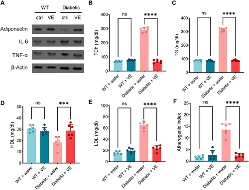
Figure 4 Vitamin E (VE) reduces maternal oxidative stress in pregnant gestational diabetes mellitus (GDM) mice at the late stage of pregnancy. The maternal serum and liver tissue were harvested on GD19. The serum (A) and liver (B) malondialdehyde (MDA) contents were measured by ELISA. The serum MDA, superoxide dismutase (SOD) (C), glutathione peroxidase (GPx) (D), glutathione (GSH) (E), and catalase (CAT) (F) in the liver were measured by ELISA. (n=6 mice). Data are presented as mean ± SD. **p < 0.01, *** p < 0.001, ****p < 0.0001.
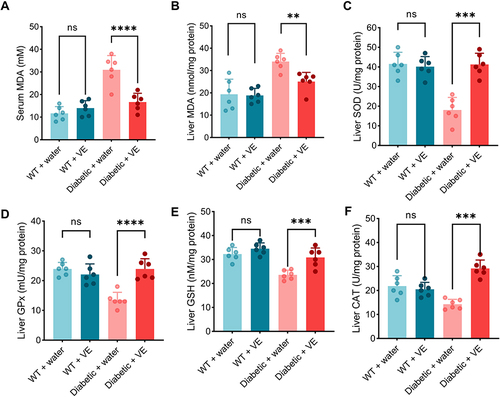
Figure 5 Vitamin E (VE) alleviates gestational diabetes mellitus (GDM) reproductive outcomes. Litter size (A) and body weight at birth (B) of the pups in different experimental groups. (n=10 pregnant mice). One data point in panel (B) represents the averaged birth weight of the pups in the litter from one mouse. Data are presented as mean ± SD. Data are presented as mean ± SD. **p < 0.01, ****p < 0.0001, ns indicates no significance.
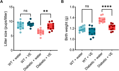
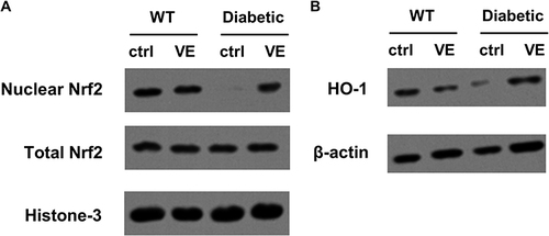
Figure 6 Effects of vitamin E (VE) on nuclear factor-erythroid factor 2-related factor 2 (Nrf2) activation and heme oxygenase-1 (HO-1) expression. The maternal liver tissues were harvested on birth. (A) Western blot analysis of total and nuclear protein levels of Nrf2. (B) Western blot analysis of HO-1 protein level.