Figures & data
Table 1 Comparisons of Clinical Parameters Among the Three Groups [n (%), (), M (P25, P75)]
Table 2 Relationship Between miR-222-3p Expression Levels in Peripheral Blood and Clinical Characteristics of DFU [n (%)]
Table 3 Relationship Between miR-222-3p Expression Levels in Wound Margin Tissue and Clinical Characteristics of DFU [n (%)]
Figure 1 Kaplan-Meier survival curve analysis evaluated the complete wound healing rate of DFU between two expression groups. (A) Group with high miR-222-3p expression presented higher unhealed DFU cumulative rate relative to the low expression group in DFU patients’ peripheral blood (log rank, P = 0.011). The estimated median time of complete wound healing in groups with high and low miR-222-3p expressions were 9.64 weeks and 8.70 weeks, respectively, in DFU patients’ peripheral blood (P < 0.05). (B) Group with high miR-222-3p expression presented higher unhealed DFU cumulative rate relative to the low expression group in DFU patients’ wound margin tissue (log rank, P = 0.001). The estimated median time of complete wound healing in high and low expression groups were 9.76 weeks and 8.71 weeks, respectively, in DFU patients’ wound margin tissue (P < 0.05).
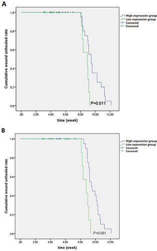
Table 4 Correlations Between miR-222-3p Expression in Peripheral Blood and Other Clinical Parameters in NC Group, T2DM Group, and DFU Group (r)
Table 5 Correlations Between miR-222-3p Expression in Wound Margin Tissue and Other Clinical Parameters in the DFU Group (r)
Table 6 The Multivariate Stepwise Logistic Regression Analysis of Risk Factors for Diabetic Foot Ulcer
Figure 2 The biomarker potential of circulating miR-222-3p for DFU and control. ROC analysis assisted in evaluating the ability of circulating miR-222-3p to distinguish between two groups. The AUC of P-miR-222-3p specific to DFU diagnosis reached 0.803 (95% CI 0.713–0.884, P < 0.001), with the optimal cut-off point of 2.52, the sensitivity of 95.93%, and the specificity of 96.27%.
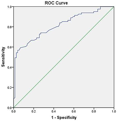
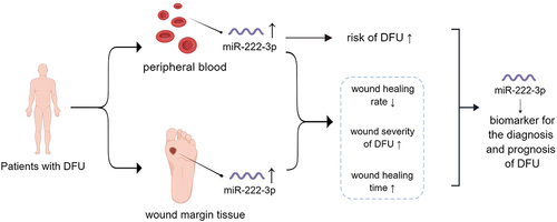
Figure 3 MiR-222-3p effectively served for DFU diagnosis and prognosis as a useful biomarker. ↑: increase; ↓: decrease.