Figures & data
Figure 1 Flow chart of patient selection.
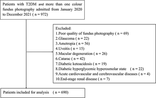
Table 1 Clinical and Laboratory Characteristics of Participants Between Non-DKD Group and DKD Group
Table 2 Comparison of the Retinal Microvascular Diameters Between Non-DKD Group and DKD Group
Table 3 The Logistic Regression Analyses Between Retinal Venular Diameters and DKD
Table 4 ORs (and 95% CIs) of DKD According to Quartiles of Retinal Venular Diameter Levelsa
Figure 2 Restricted cubic splines for the association between retinal venular diameters and DKD. OR of DKD by the diameters of superior temporal retinal venula (A), inferior temporal retinal venula (B), superior nasal retinal venula (C), inferior nasal retinal venula (D) and CRVE (E) with the use of restricted cubic splines. The analysis was adjusted for duration of T2DM, hypertension, SBP, DBP, BMI, TG, HDL-C, BUN, Scr and UA.
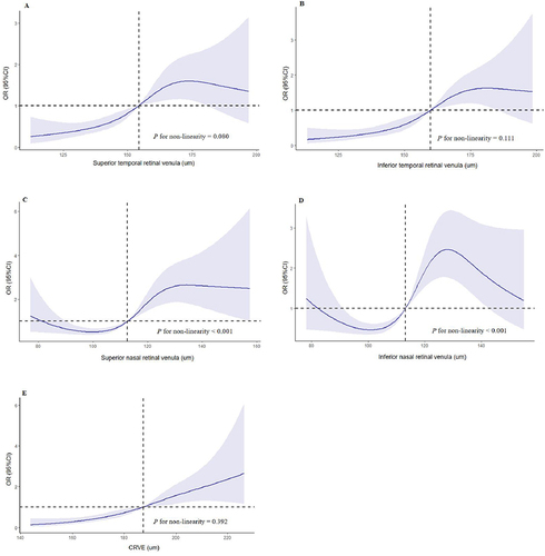
Table 5 The Logistic Regression Analyses Between Retinal Arteriolar Diameters and DKD
Table 6 ORs (and 95% CIs) of DKD According to Quartiles of Retinal Arteriolar Diameter Levelsa
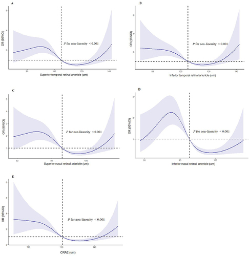
Figure 3 Restricted cubic splines for the association between retinal arteriolar diameters and DKD. OR of DKD by the diameters of superior temporal retinal arteriole (A), inferior temporal retinal arteriole (B), superior nasal retinal arteriole (C), inferior nasal retinal arteriole (D) and CRAE (E) with the use of restricted cubic splines. The analysis was adjusted for duration of T2DM, hypertension, SBP, DBP, BMI, TG, HDL-C, BUN, Scr and UA.