Figures & data
Table 1 Information for Antibodies Used in This Study
Figure 1 Effect of let-7b-5p on the lipid accumulation of C2C12 myotube cells. (A) The relative expression of let-7b-5p in TC-tet, KPC, and Pan02 cells; (B) Western blotting results of the expression of CD9 and CD63 expressed in the isolated PC cell-derived exosomes; (C) qRT-PCR detection of let-7b-5p in the isolated PC cell-derived exosomes; (D) The relative expression of let-7b-5p in C2C12 myotube cells after let-7b-5p inhibitor transfection for 24h or 48h; (E) Oil red O straining results of the accumulation of C2C12 myotube cells transfected with let-7b-5p inhibitor with PC cells-derived exosomes, the red part indicates the stained lipid particles. *P < 0.05; **P<0.01.
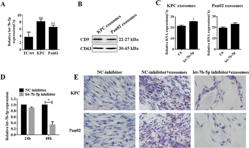
Figure 2 Exosomal let-7b-5p exerted its role on IR by activating the STAT3/FOXO1 pathway. (A and B) Western blotting results of the expression of IRS-1, GLUT4, STAT3, p-STAT3, total protein FOXO1 and nuclear FOXO1 in C2C12 myotube cells transfected with let-7b-5p inhibitor with PC cells-derived exosomes; the histograms indicated the quantification and statistical analysis of protein bands, β-actin was used as a control for total protein, and H3 was used as a control of nuclear proteins; (C&D) The relative mRNA expression of STAT3 of C2C12 myotube cells co-incubated with KPC exosomes (C) and Pan02 exosomes (D) after treated with NC-inhibitor or let-7b-5p inhibitor. *P < 0.05, **P < 0.01.
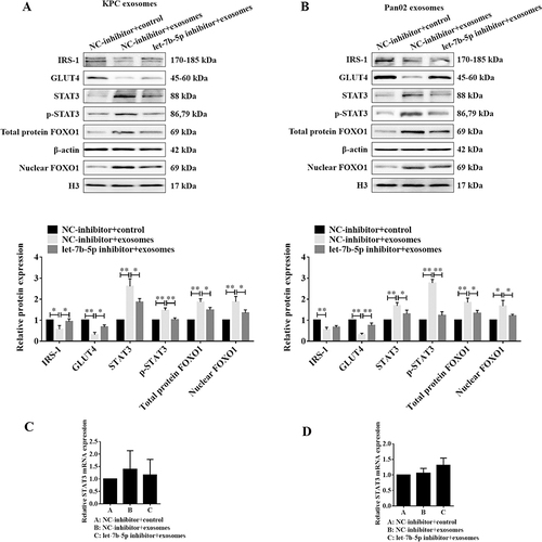
Figure 3 Let-7b-5p inhibitor reverses the exosome-induced decrease of RNF20 expression in C2C12 myotube cells. (A–D) Western blotting results of RNF20 expression in C2C12 myotube cells co-cultured with KPC exosomes (left) or Pan02 exosomes (right) after treatment with let-7b-5p inhibitor and quantification of the protein bands, and β-actin was used as a control; (E–F) qRT-PCR detection of RNF20 mRNA expression in C2C12 myotube cells treated as in A and B. *P < 0.05, **P < 0.01.
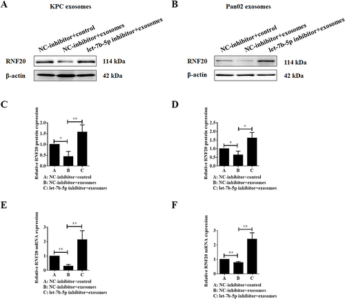
Figure 4 Validation of the targeting relationship between let-7b-5p and RNF20. (A) qRT-PCR assay was performed to validate the transfection efficiency of let-7b-5p mimics in C2C12 myotube cells; (B) Western blotting results of RNF20 expression in C2C12 myotube cells transfected with let-7b-5p mimics or NC for 24h, and quantification of the protein bands, and β-actin was used as a control; (C) qRT-PCR assay data RNF20 mRNA expression in C2C12 myotube cells treated as in A; (D) TargetScan Human 8.0 (https://www.targetscan.org) was used for predicting the targeting relationship and binding sites between let-7b-5p and RNF20; (E) Dual-luciferase activity reporter assay results of in C2C12 myotube cells co-transfected with let-7b-5p mimics and luciferase reporter plasmids containing wildtype or mutant RNF20 3’UTR; (F–G) Western blotting results of RFN20 overexpression (F), and quantification of protein band, β-actin was used as a control (G); (H and I) IP/WB experiment was used to detect the ubiquitination and expression of STAT3 in C2C12 myotube cells co-transfected with let-7b-5p mimics and/or RFN20 overexpression plasmid (H), and quantification of protein band, β-actin was used as a control (I). *P<0.05, **P<0.01.
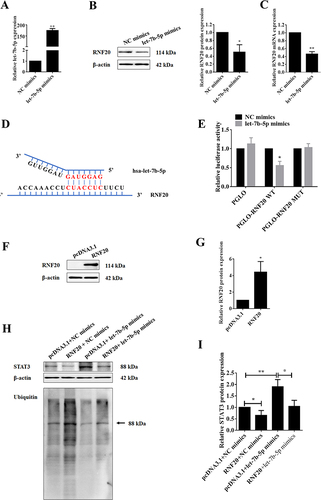
Figure 5 Overexpression of RNF20 reverses PC cell-derived exosome-induced insulin resistance. (A) Oil Red O staining assay of lipid accumulation in C2C12 myotube cells treated with exosomes or co-overexpressed RFN20, the red part indicates the stained lipid particles. (B) Western blotting results of the expression of RNF20, IRS-1, GLUT4, STAT3, p-STAT3, total protein FOXO1 and nuclear FOXO1; (C) quantitative analysis of protein band, β-actin was used as a control of total protein. *P<0.05, **P<0.01.
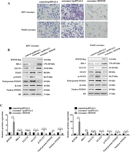
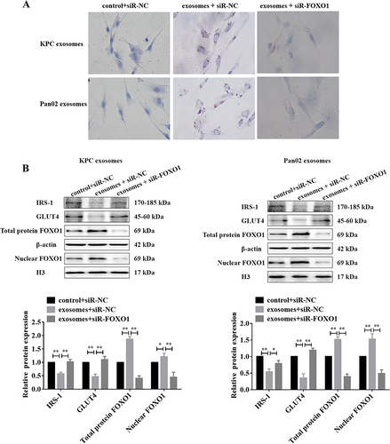
Figure 6 Knocking FOXO1 down reverses PC cell-derived exosome-induced insulin resistance. (A) Oil Red O staining assay of lipid accumulation in C2C12 myotube cells treated with exosomes or co-silenced FOXO1, the red part indicates the stained lipid particles. (B) Western blotting results of the expression of IRS-1, GLUT4, total protein FOXO1 and nuclear FOXO1; and the quantitative results of the immunoblots were shown in the histogram below. β-actin was used as a control of total protein. *P<0.05, **P<0.01.