Figures & data
Figure 1 Iron enters the brain and is transported to neurons and glial cells. Iron-binding Tf binds to TfR and is internalized in endosomes. Acidification of endosomes dissociates Fe from Tf and reduces it to ferrous ions, which are subsequently released into the cytoplasm by DMT1. Finally, ferrous ions leave BMVECs through FPN1 and are exported into the interstitial fluid of the brain, where they are oxidized to ferric by ceruloplasmin. Neurons and microglia mainly take up Fe through the Tf-Tfr1 system, while astrocytes can take up iron directly from BMVECs through DMT1. Created with Biorender.com.
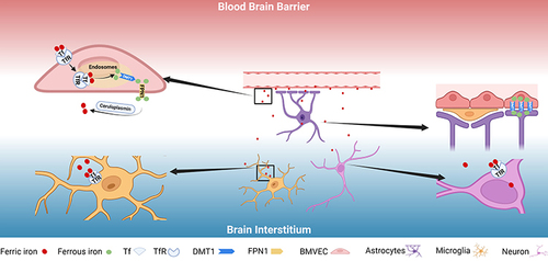
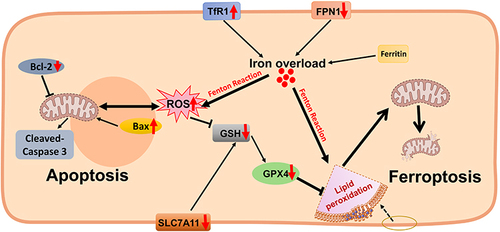
Figure 2 Excess iron-induced cell death in the diabetic brain. The expression of TfR1 and DMT1 is up-regulated in the diabetic brain (especially in hippocampal neurons), and the expression of FPN1 is down-regulated, which may lead to iron overload. As a direct consequence, iron overload can increase ROS and lipid oxide levels and reduce antioxidant activity such as GSH through the Fenton reaction. On the one hand, this leads to the damage of mitochondrial structure and function, the down-regulation of GPX4 and SLC7A11 protein expression and triggers neuronal ferroptosis; on the other hand, it may lead to the up-regulation of Bax protein and the down-regulation of Bcl-2 protein, triggering apoptosis. Some of the materials in are created with Biorender.com.