Figures & data
Figure 1 Enrollment flowchart of the participants.
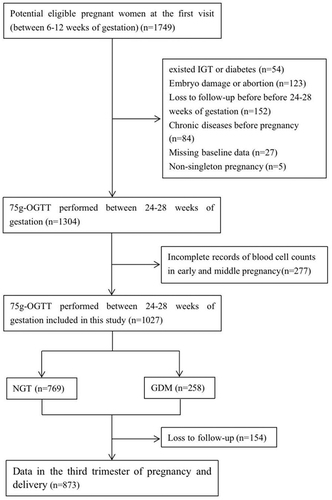
Table 1 Characteristics and Laboratory Data of the Study Participants
Figure 2 The changes of WBC (a), RBC (b), HGB (c), and PLT (d) from early to middle pregnancy in the non-GDM and GDM group. Data were demonstrated by mean and 95% CI.
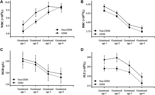
Table 2 The Odds Ratios (ORs) of GDM Across Increased Quartiles of Hematological Parameters During Different Gestational Ages
Figure 3 The forest plots of subgroup analyses of WBC (a), RBC (b), and PLT (c) at gestational age 1 and 2 by maternal age and preBMI. The ORs were adjusted for maternal age, preBMI, family history of diabetes, and history of adverse pregnancy (defined as embryo damage, spontaneous abortion or preterm delivery). *P<0.05 and **P<0.01.
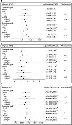
Figure 4 The association between WBC, RBC, and PLT at different gestational ages and FINS in the first trimester.

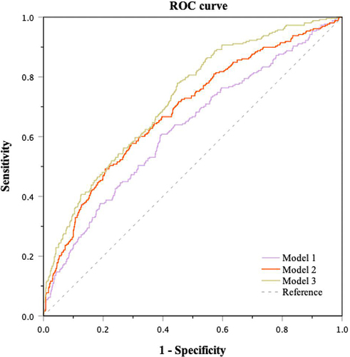
Figure 5 The ability of blood cell counts in early pregnancy combined with other parameters for the prediction of GDM. Model 1: WBC, RBC, and PLT at gestational age 1 and 2; Model 2: Model 1 + maternal age, preBMI, family history of diabetes, and history of adverse pregnancy; Model 3: Model 2 + FBG and FINS in the first trimester.