Figures & data
Figure 1 Chemical structure of streptozotocin.
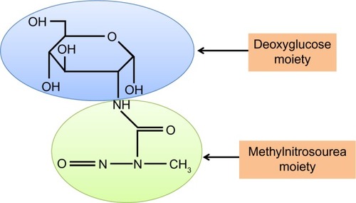
Figure 2 Mechanisms by which diabetic hyperglycemia can impose glucotoxicity on β cells.
Notes: The biochemical pathways listed can be upregulated or activated by chronic hyperglycemia. Importantly, all these pathways are eventually involved in elevated production of reactive oxygen species that are detrimental to β cell function. These pathways can be dissected by animal models of STZ diabetes.
Abbreviations: STZ, streptozotocin; ROS, reactive oxygen species.
Abbreviations: STZ, streptozotocin; ROS, reactive oxygen species.
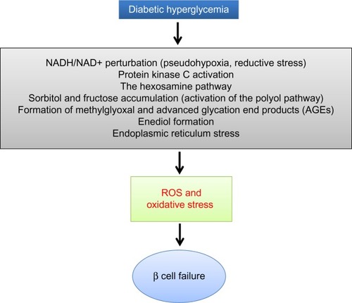
Figure 3 Glucose combustion is tightly coupled to insulin secretion in pancreatic β cells.
Notes: The figure shown depicts the main pathways of glucose metabolism and mitochondrial ATP production. Glucose is first transported into β cells via GLUT2 transporters, followed by glycolysis, Krebs cycle, and oxidative phosphorylation that eventually make ATP from the combustion of glucose. The elevated ratio of ATP/ADP, driven by high blood glucose, closes the KATP channel and opens the calcium channel on the cell membranes. The influx of calcium triggers exocytosis of insulin granules and subsequent insulin release.
Abbreviation: TCA, tricarboxylic acid.
Abbreviation: TCA, tricarboxylic acid.
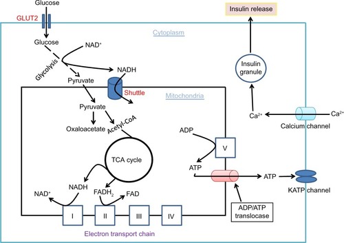
Figure 4 Role of redox imbalance between NADH and NAD+ in β cell dysfunction.
Notes: Under euglycemic condition, the balance between NADH and NAD+ is well maintained. However, under diabetic hyperglycemic condition, the balance between NADH and NAD+ is broken by several mechanisms such as NADH overproduction via the glycolytic and the polyol pathways and NAD+ depletion by poly ADP-ribosylase, sirtuins, and CD38. Albeit intensive studies in the field of diabetes, the role of complex I that makes NAD+ from NADH in this redox imbalance is unknown, so is the role of those enzymes making NADH from NAD+.
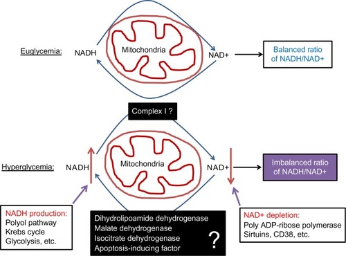
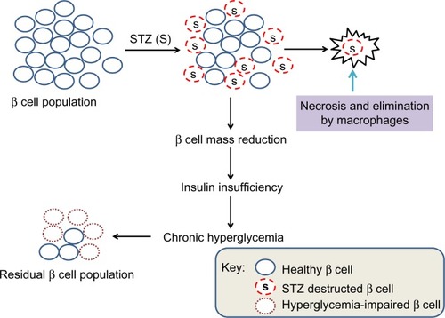
Figure 5 Scheme showing partial destruction of β cell population by STZ and reduction in β cell mass that induces insulin insufficiency and chronic hyperglycemia.
Notes: While STZ-destructed β cells undergo necrosis and elimination by macrophages, the surviving or residual β cells are exposed to persistent hyperglycemia that can impair mitochondrial function in the residual β cell population.
Abbreviation: STZ, streptozotocin.
Abbreviation: STZ, streptozotocin.