Figures & data
Table 1 Bodyweight and mean arterial blood pressure, blood glucose, and percentage HbA1c
Figure 1 Co-immunofluorescent staining of protein kinase C (PKC)-βII and vascular smooth muscle cells in mesenteric arteries (diameter =100–120 μm) taken from control, diabetes mellitus (DM), and DM curcumin 300 (DM-CUR 300) groups. Double-immunofluorescent staining was performed to demonstrate PKC-βII in the mesenteric arteries (red fluorescence; A), α-smooth muscle actin (green fluorescence; B), and merged image (yellow; C). Magnification: ×400.
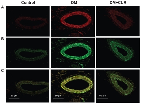
Figure 2 A) Immunostaining for cyclo-oxygenase (COX)-2 in small mesenteric arteries. Sections of arterial segment show the endothelial cell layer inwards. Positive immunoreactions are observed as a brown precipitate.
Abbreviations: CON-NSS, control with 0.9% normal saline; DM-CUR30, diabetes-treated with curcumin 30 mg/kg; DM-CUR300, diabetes-treated with curcumin 300 mg/kg; DM-NSS, diabetes treated with 0.9% normal saline; SEM, standard error of the mean.
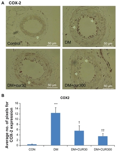
Figure 3 A) The expression of nuclear factor-κB p65 (NF-κB p65) in small mesenteric arteries in the control (left upper panel), DM (right upper panel), DM-CUR30 (left lower panel) and DM-CUR300 (right lower panel) rats. Positive immunoreactions are observed as a brown precipitate. Magnification: ×400. B) Expression of NF-κB in the endothelium layer in small mesenteric arteries using image analysis (Global Lab Image/2 software) measurement in the DM-NSS, DM-CUR30, and DM-CUR300 groups.
Abbreviations: DM-CUR30, diabetes-treated with curcumin 30 mg/kg; DM-CUR300, diabetes-treated with curcumin 300 mg/kg; DM-NSS, diabetes treated with 0.9% normal saline; NF-κB, nuclear factor-κB; SEM, standard error of the mean.
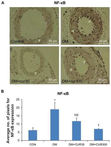
Table 2 Means ±SEM of basal levels of 6-keto-PGF1α and TXB2 together with the 6-keto-PGF1α/TXB2 ratios in the mesenteric arteriolar bed of CON-NSS, DM-NSS, and DM-CUR300 groups
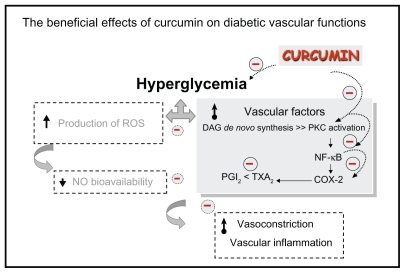
Figure 4 The beneficial effects of curcumin on diabetic vascular functions. Curcumin supplementation (300 mg/kg bodyweight) improved diabetes-induced vascular dysfunction associated with its potential to reduce blood sugar, COX-2 and NF-κB suppression, PKC inhibition, and improve the ratio of prostanoid products PGI2 and TXA2.