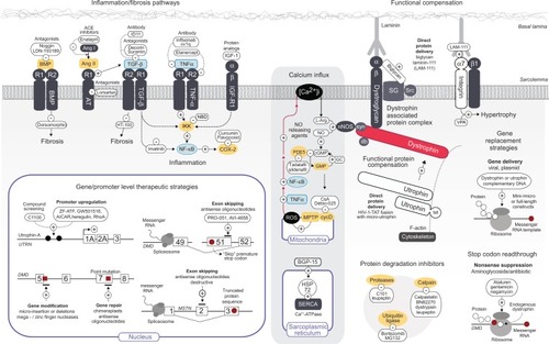
Figure 1 Current genetic and pharmacological targets of dystrophic pathology.
Notes: Receptor or structural protein components at the skeletal muscle sarcolemma targeted for therapeutic purposes are represented in dark grey. Components of signaling pathways specifically targeted for intentional downregulation are represented in yellow boxes, with two key regulators of dystrophic pathology NFκB, and TNFα, highlighted in light blue. Associated white boxes contain the therapeutic compound(s) used to modulate either a positive (+) or negative (−) effect on a particular protein/receptor. Arrow-headed lines represent a simplified version of the signaling pathways involved in inflammatory, fibrotic, and hypertrophic responses, symbolizing only regulatory proteins that are covered in the text. The background-shaded section represents cellular process affected by calcium influx, with the red line representing the feedback mechanism with ROS, TNFα, and NFκB. The interactions delineated between and within signaling pathways are not an exhaustive representation and pharmacological compounds that act in a nonspecific or undetermined mode of action have been excluded. Red dots, representative mRNA sequence leading to translation stop codon; black dots, promoter elements; red squares, representative of gene modification by repair or insertion/deletion by zinc finger or meganucleases. Abbreviations: PA, polyaxamer 188; TGF-β, transforming growth factor beta; PI3K, phosphatidylinositol 3-kinase; AKT, protein kinase B (PKB); IKK, IκB kinase; NF-κB, nuclear factor kappa-light-chain enhancer of activated B cells; BMP, bone morphogenic protein; AngII, angiotensin II; RI/RII, receptor I/II; AT, angiotensin; ACE, angiotensin-converting enzyme; NBD, NEMO binding domain; IGF-1, insulin growth factor 1; LAM-III, laminin-111 protein; VPA, valproic acid; ALK-4, activin receptor-like kinase; ActRII, activin receptor type II; BCL/ABL, breakpoint cluster region/Abelson murine leukemia viral oncogene homologue 1; Akt, acutely transforming retrovirus AKT8 in rodent T-cell lymphoma; [Ca2+]i, intracellular calcium; L-Arg, L-arginine; NO, nitric oxide; nNOS, neural nitric oxide synthase; cGMP, cyclic guanosine monophosphate; GMP, guanosine monophosphate; GC, guanylate cyclase; BGP-15, O-(3-piperidino-2-hydroxy-1-propyl) nicotinic amidoxime; PDE5, cGMP-specific phosphodiesterase type 5; TNF-α, tumour necrosis factor alpha; CsA, cyclosporine A; ROS, reactive oxygen species; MPTP, mitochondrial permeability transition pore; cycD, cyclophilin D; HSP72, heat shock protein 72; SERCA, sarco/endoplasmic reticulum Ca2+-ATPase; SG, sarcoglycans; Src, sarcospan; syn, sytrophin; db, dystrobrevin; RDO, RNA/DNA oligonucleotide; AON, antisense oligonucleotide; 2′OMePS, 2′-O-methyl oligoribonucleotide; PMO, phosphorodiamidate morpholino oligomer; AICAR, 5-amino-1-β-D-ribofuranosyl-imidazole-4-carboxamide; UTRN/DMD/MSTN, utrophin/DMD/myostatin gene; DAPC, dystrophin-associated protein complex; LAM, laminin; Utr, utrophin minigene construct; cDNA, complementary DNA.
BulfieldGSillerWGWightPAMooreKJX chromosome-linked muscular dystrophy (mdx) in the mouseProc Natl Acad Sci U S A1984814118911926583703
SicinskiPGengYRyder-CookASBarnardEADarlisonMGBarnardPJThe molecular basis of muscular dystrophy in the mdx mouse: a point mutationScience19892444912157815802662404
MoissetPASkukDAsselinISuccessful transplantation of genetically corrected DMD myoblasts following ex vivo transduction with the dystrophin minigeneBiochem Biophys Res Commun1998247194999636661
MoissetPAGagnonYKarpatiGTremblayJPExpression of human dystrophin following the transplantation of genetically modified mdx myoblastsGene Ther1998510134013469930339
SonnemannKJHeun-JohnsonHTurnerAJBaltgalvisKALoweDAErvastiJMFunctional substitution by TAT-utrophin in dystrophin-deficient micePLoS Med200965e100008319478831
AmentaARYilmazABogdanovichSBiglycan recruits utrophin to the sarcolemma and counters dystrophic pathology in mdx miceProc Natl Acad Sci U S A2010108276276721187385
RooneyJEGurpurPBBurkinDJLaminin-111 protein therapy prevents muscle disease in the mdx mouse model for Duchenne muscular dystrophyProc Natl Acad Sci U S A2009106197991799619416897
RooneyJEGurpurPBYablonka-ReuveniZBurkinDJLaminin-111 restores regenerative capacity in a mouse model for alpha7 integrin congenital myopathyAm J Pathol2009174125626419074617
BertoniCRandoTADystrophin gene repair in mdx muscle precursor cells in vitro and in vivo mediated by RNA-DNA chimeric oligonucleotidesHum Gene Ther200213670771811936970
Barton-DavisERCordierLShoturmaDILelandSESweeneyHLAminoglycoside antibiotics restore dystrophin function to skeletal muscles of mdx miceJ Clin Invest1999104437538110449429
LoufraniLDubrocaCYouDAbsence of dystrophin in mice reduces NO-dependent vascular function and vascular density: total recovery after a treatment with the aminoglycoside gentamicinArterioscler Thromb Vasc Biol200424467167614751810
GurpurPBLiuJBurkinDJKaufmanSJValproic acid activates the PI3K/Akt/mTOR pathway in muscle and ameliorates pathology in a mouse model of Duchenne muscular dystrophyAm J Pathol20091743999100819179609
BodineSCStittTNGonzalezMAkt/mTOR pathway is a crucial regulator of skeletal muscle hypertrophy and can prevent muscle atrophy in vivoNat Cell Biol20013111014101911715023
AsaiASahaniNKanekiMOuchiYMartynJAYasuharaSEPrimary role of functional ischemia, quantitative evidence for the two-hit mechanism, and phosphodiesterase-5 inhibitor therapy in mouse muscular dystrophyPLoS One200728e80617726536
AdamoCMDaiDFPercivalJMSildenafil reverses cardiac dysfunction in the mdx mouse model of Duchenne muscular dystrophyProc Natl Acad Sci U S A201010744190791908320956307
FaircloughRJPerkinsKJDaviesKEPharmacologically targeting the primary defect and downstream pathology in duchenne muscular dystrophyCurr Gene Ther201212320624422571500
ChapmanVMMillerDRArmstrongDCaskeyCTRecovery of induced mutations for X chromosome-linked muscular dystrophy in miceProc Natl Acad Sci U S A1989864129212962919177
KimuraELiSGregorevicPFallBMChamberlainJSDystrophin delivery to muscles of mdx mice using lentiviral vectors leads to myogenic progenitor targeting and stable gene expressionMol Ther201018120621319888194
MitrpantCFletcherSIversenPLWiltonSDBy-passing the nonsense mutation in the 4 CV mouse model of muscular dystrophy by induced exon skippingJ Gene Med2009111465619006096
BachrachELiSPerezALSystemic delivery of human microdystrophin to regenerating mouse dystrophic muscle by muscle progenitor cellsProc Natl Acad Sci U S A2004101103581358614993597
MaguireKSuzukiTDiMatteoDParekh-OlmedoHKmiecEGenetic correction of splice site mutation in purified and enriched myoblasts isolated from mdx5cv miceBMC Mol Biol2009101519236710
HibaouiYReutenauer-PatteJPatthey-VuadensORueggUTDorchiesOMMelatonin improves muscle function of the dystrophic mdx5Cv mouse, a model for Duchenne muscular dystrophyJ Pineal Res201151216317121486366
ArakiENakamuraKNakaoKTargeted disruption of exon 52 in the mouse dystrophin gene induced muscle degeneration similar to that observed in Duchenne muscular dystrophyBiochem Biophys Res Commun199723824924979299538
AokiYNakamuraAYokotaTIn-frame dystrophin following exon 51-skipping improves muscle pathology and function in the exon 52-deficient mdx mouseMol Ther201018111995200520823833
BurkinDJWallaceGQNicolKJKaufmanDJKaufmanSJEnhanced expression of the alpha 7 beta 1 integrin reduces muscular dystrophy and restores viability in dystrophic miceJ Cell Biol200115261207121811257121
BerrySELiuJChaneyEJKaufmanSJMultipotential mesoangioblast stem cell therapy in the mdx/utrn−/− mouse model for Duchenne muscular dystrophyRegen Med20072327528817511564
Rafael-FortneyJAChimanjiNSSchillKEEarly treatment with lisinopril and spironolactone preserves cardiac and skeletal muscle in Duchenne muscular dystrophy miceCirculation2011124558258821768542
ShiSHoogaarsWMde GorterDJBMP antagonists enhance myogenic differentiation and ameliorate the dystrophic phenotype in a DMD mouse modelNeurobiol Dis201141235336020940052
PetersonJMKlineWCananBDPeptide-based inhibition of NF-kappaB rescues diaphragm muscle contractile dysfunction in a murine model of Duchenne muscular dystrophyMol Med2011175–650851521267511
GehrigSMvan der PoelCHoeflichANaimTLynchGSMetzgerFTherapeutic potential of PEGylated insulin-like growth factor I for skeletal muscle disease evaluated in two murine models of muscular dystrophyGrowth Horm IGF Res2012222697522424862
Bremmer-BoutMAartsma-RusAde MeijerEJTargeted exon skipping in transgenic hDMD mice: a model for direct preclinical screening of human-specific antisense oligonucleotidesMol Ther200410223224015294170
Arechavala-GomezaVGrahamIRPopplewellLJComparative analysis of antisense oligonucleotide sequences for targeted skipping of exon 51 during dystrophin pre-mRNA splicing in human muscleHum Gene Ther200718979881017767400
GoyenvalleAWrightJBabbsAWilkinsVGarciaLDaviesKEEngineering multiple U7snRNA constructs to induce single and multiexon-skipping for Duchenne muscular dystrophyMol Ther20122061212122122354379
SharpNJKornegayJNVan CampSDAn error in dystrophin mRNA processing in golden retriever muscular dystrophy, an animal homologue of Duchenne muscular dystrophyGenomics19921311151211577476
KornegayJNLiJBoganJRWidespread muscle expression of an AAV9 human mini-dystrophin vector after intravenous injection in neonatal dystrophin-deficient dogsMol Ther20101881501150820517298
KooTMalerbaAAthanasopoulosTDelivery of AAV2/9-microdystrophin genes incorporating helix 1 of the coiled-coil motif in the C-terminal domain of dystrophin improves muscle pathology and restores the level of alpha1-syntrophin and alpha-dystrobrevin in skeletal muscles of mdx miceHum Gene Ther201122111379138821453126
BartlettRJStockingerSDenisMMIn vivo targeted repair of a point mutation in the canine dystrophin gene by a chimeric RNA/DNA oligonucleotideNat Biotechnol200018661562210835598
BishLTSleeperMMForbesSCLong-term restoration of cardiac dystrophin expression in golden retriever muscular dystrophy following rAAV6-mediated exon skippingMol Ther201220358058922146342
BarbashIMCecchiniSFaraneshAZMRI roadmap-guided transendocardial delivery of exon-skipping recombinant adeno-associated virus restores dystrophin expression in a canine model of Duchenne muscular dystrophyGene Ther Epub532012
TownsendDTurnerIYasudaSChronic administration of membrane sealant prevents severe cardiac injury and ventricular dilatation in dystrophic dogsJ Clin Invest201012041140115020234088
ChildersMKBoganJRBoganDJChronic administration of a leupeptin-derived calpain inhibitor fails to ameliorate severe muscle pathology in a canine model of duchenne muscular dystrophyFront Pharmacol201128922291646
OhshimaSShinJHYuasaKTransduction efficiency and immune response associated with the administration of AAV8 vector into dog skeletal muscleMol Ther2009171738018941441
YokotaTHoffmanETakedaSAntisense oligo-mediated multiple exon skipping in a dog model of duchenne muscular dystrophyMethods Mol Biol201170929931221194037
Nitahara-KasaharaYHayashita-KinohHOhshima-HosoyamaSLong-term engraftment of multipotent mesenchymal stromal cells that differentiate to form myogenic cells in dogs with Duchenne muscular dystrophyMol Ther201220116817721934652
WalmsleyGLArechavala-GomezaVFernandez-FuenteMA duchenne muscular dystrophy gene hot spot mutation in dystrophin-deficient cavalier king charles spaniels is amenable to exon 51 skippingPLoS One201051e864720072625
WinandNCooperBMolecular characterization of severe Duchenne-type muscular dystrophy in a family of rottweiler dogsProceedings of Molecular Mechanisms of Neuromuscular DiseaseTucsonUniversity of Arizona1994