Figures & data
Figure 1 Selected photophobia neural pathways in dry eye and migraine. Light evokes signals from rod and cone cells that are transmitted via amacrine and bipolar cells (not shown) to retinal ganglion cells (RGC), which project to the olivary pretectal nucleus (OPN, green line). Blue line: parasympathetic signals travel from the OPN to the superior salivatory nucleus (SSN), then to the sphenopalatine ganglion (SPG), and ocular and dural vessels to mediate vasodilation. Red line: afferent trigeminal signals from cornea (stimulated by corneal disruptions), ocular vessels, and dural vessels (stimulated by vasodilation) travel to the trigeminal ganglion (TG) then to the trigeminal nucleus caudalis (TNC) and finally the posterior thalamus. Alternatively, light-evoked signals from intrinsically photosensitive RGCs (ipRGC) travel directly to the posterior thalamus (purple line). Black line: signals from the posterior thalamus travel to somatosensory and visual cortices to mediate dry eye and migraine symptoms. Note other pathways of photophobia that involve the hypothalamus and retinal rod and cone cells are not depicted.
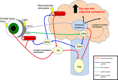
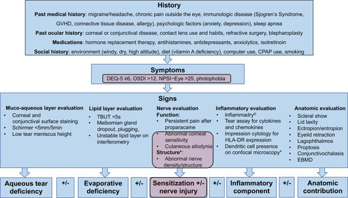
Figure 2 Clinical assessment of patients with dry eye. Purple boxes indicate phenotypes that overlap with migraine. *Nerve structure findings using in-vivo confocal microscopy.