Figures & data
Figure 1 Ultrawide field scanning laser ophthalmoscope (Optomap®) image.
Note: Areas 1–9 are delineated to denote location of lesions during image review (Copyright Optos, All rights reserved, reprinted with permission).
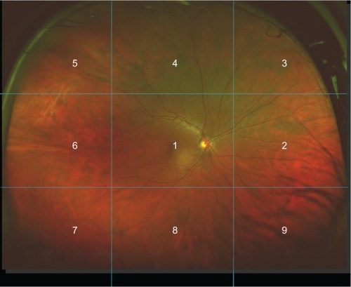
Image 1 Nonmydriatic, ultrawide field image of a peripheral retinal hemorrhage adjacent to an area of retinal thinning (area in circle) in a 26-year-old, healthy, asymptomatic subject.
Notes: The insert is a green laser channel view of the same peripheral hemorrhage. Small yellow artifact at center of image and eyelashes at lower edge of image. Brightness and contrast of image have been enhanced to increase visibility of lesion.
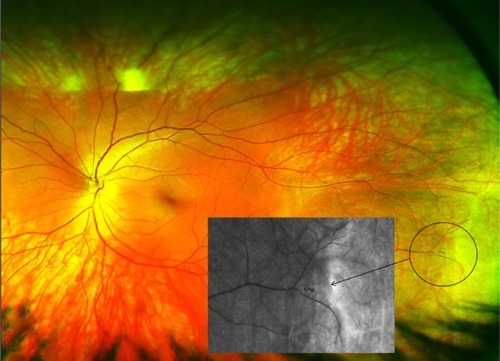
Table 1 Prevalence of lesions (n = 170, 339 eyes)Table Footnote*
Table 2 Exact agreement between traditional and image-assisted ophthalmoscopy
Table 3 Proportion of observations agreeing with adjudication (n = 170, 339 eyes)Table Footnote*
Table 4 Evaluation of discordant results