Figures & data
Table 1 Key Terms in Ocular Motor Research, Examinations and Analysis and Their Definitions
Figure 1 Schematic representation of saccadic pathways. Voluntary saccades arise from frontal eye fields and travel to the basal ganglia (caudate nucleus, substantia nigra) before descending to the superior colliculus and forward onto brainstem eye movement structures. Additional brain regions (egDLPFC) may be recruited for to execute more cognitively complex saccades, such as anti-saccades. Reflexive saccades have a direct pathway from visual cortex (not shown) to the posterior parietal cortex and from here onto the superior colliculus. There is an additional pathway from the posterior parietal cortex to activate frontal eye fields and then travel via the basal ganglia towards the superior colliculus, and reticular formation before entering the midbrain (vertical) and pontine (horizontal) ocular motor plant. dlPFC dorsolateral prefrontal cortex.
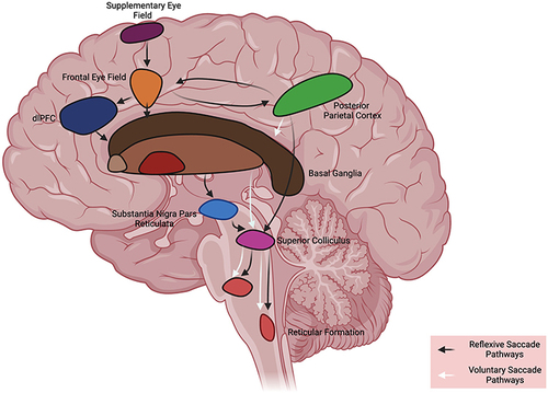
Table 2 Quantitative Metrics Acquired from Classes of Eye Movements
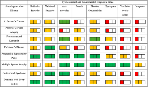
Figure 2 Ocular motor features of neurodegenerative diseases and their associated diagnostic value (red: low diagnostic value, yellow: medium diagnostic value and green: high diagnostic value) based on key publications highlighting ocular motor features of each neurodegenerative disease.