
Figures & data
Figure 1 External photograph demonstrating left Horner’s syndrome secondary to a left neck tumor in a 62-year-old woman. Note left ptosis in daylight (figure at the top) and left miosis in the left eye on the right more pronounced in dim light (figure at bottom). An informed written consent was obtained from the patient.
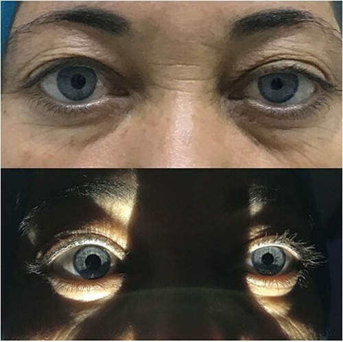
Figure 2 Right congenital Horner’s syndrome in a 10-year-old girl. Right ptosis, iris hypochromia and anisocoria in dim light can be seen (right eye on the left). An informed written consent was obtained from the patient.
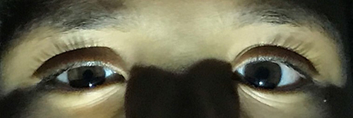
Figure 3 A 42-year-old woman presented with a 6-month history of persistent headache associated with a posterior neck pain that recently worsened. Right Horner’s syndrome was found on examination with right ptosis, anisocoria and conjunctival hyperemia (right eye on the left). CT-scan revealed the presence of bilateral Eagle’s syndrome, which was more severe on the right side. CBH occurrence is explained by a compression along the oculosympathetic pathway by the abnormal elongation of the styloid process. An informed written consent was obtained from the patient. Reprinted from Maamouri R, Ouederni M, Oueslati Y, Mbarek C, Chammakhi C and Cheour M. Acute Painful Horner’s Syndrome Revealing Eagle’s Syndrome: A Report of Two Cases. Neuro-Ophthalmology. 2022;46(4), 244–247.Citation25
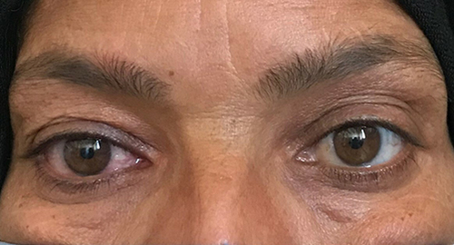
Table 1 Etiologies of Horner’s Syndrome According to the Site of the Lesion