Figures & data
Figure 1 Neurophysiological measures within a cortical volume.
Notes: (A) In most neurophysiological preparations, microelectrodes are inserted into the gray matter and time-varying voltage is measured against a nearby reference. (B) If recordings are performed at more than one location simultaneously, one can compare signals along the radial dimension across the cortical layers or along the tangential dimension that runs in parallel with the cortical surface. While radial recordings are often performed with linear multielectrode arrays that consist of multiple electrode contacts that run along a single electrode shaft, tangential recordings typically require two or more needle-shaped microelectrodes spaced apart.
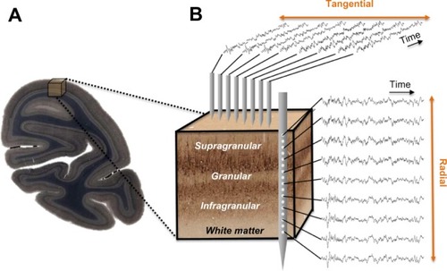
Figure 2 LFP coherence as a function of tangential and radial distance.
Notes: (A) LFP coherence (ordinate) is computed between a seed electrode in the primary visual cortex and several neighboring electrodes that are spaced at varying intervals of tangential cortical distance (abscissa). Each point of the graph depicts the LFP coherence value measured at that cortical recording location with respect to the seed position. High frequency LFP coherence (red line) and low frequency LFP coherence (blue line) are shown separately. Note the difference in tangential falloff of LFP coherence, indicating that slow neural processes are more widespread along the cortical mantle compared with locally confined fast neural activity (see Leopold et alCitation74 for details and statistics). (B) LFP coherence as a function of radial (laminar) cortical distance. Each point of the graph corresponds to an electrode contact position of a linear electrode array that was placed to record neural activity across all of primary visual cortex’s layers between the pia mater and the white mater. LFP coherence for each of these recording locations is computed against the electrode contact in the infragranular layers marked as the seed position. Dashed horizontal line marks the transition zone between the granular and infragranular layers. Note that for the same radial cortical distance LFP coherence remains higher in the infragranular compartment than in the supragranular compartment (see Maier et alCitation79 for details and statistics).
Abbreviations: LFP, local field potential; sg, supragranular; g, granular; ig, infragranular.
Abbreviations: LFP, local field potential; sg, supragranular; g, granular; ig, infragranular.
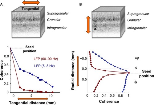
Figure 3 Volumetric profile of LFP coherence.
Notes: (A) LFP coherence is shown as a function of frequency and tangential distance relative to a seed electrode at distance 0. Note the monotonic falloff of LFP coherence with tangential cortical distance (see Leopold et alCitation74 for details). (B) LFP coherence is shown as a function of frequency and cortical depth. A seed electrode (yellow dashed line) was chosen in the supragranular layers (left) or in the infragranular layers (right), and coherence was computed as a function of frequency in spatial increments of 100 microns. Note the falloff of coherence for high frequency activity in the laminar compartment where the seed is located (see Maier et alCitation79 for details).
Abbreviations: LFP, local field potential; sg, supragranular; g, granular; ig, infragranular.
Abbreviations: LFP, local field potential; sg, supragranular; g, granular; ig, infragranular.
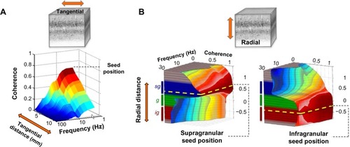
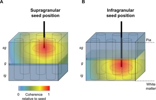
Figure 4 Schematic representation of the spatial anisotropy of LFP coherence along the tangential and radial dimensions of primate primary visual cortex.
Notes: (A) Graphic depicting the spatial falloff in LFP coherence with increasing distance from an electrode location in the supragranular layers. The three-dimensional expanse of the neocortical tissue sample is depicted as a blue-grayish box. Cortical columns are conceptualized as black cylinders. The electrode tip is indicated as “seed”. (B) Same as A, but with electrode recording location in the deep (infragranular) layers of cortex.
Abbreviations: LFP, local field potential; sg, supragranular; g, granular; ig, infragranular.
Abbreviations: LFP, local field potential; sg, supragranular; g, granular; ig, infragranular.