Figures & data
Figure 1 Computer screenshot of the interface of a telemedicine software.
Notes: A standard set of six photos is captured (external photo, posterior pole centered around the nerve, temporal, nasal, inferior, and superior). The software is capable of generating a montage of the acquired images.
Abbreviations: PMA, post-menstrual age; Cest, gestational age; SUP, superior; EXT, external; TEMP, temporal; POST, posterior pole; NAS, nasal; INF, inferior; Indet, indeterminate; S1, Stage 1; S2, Stage 2; S3 Flat, Stage 3 with flat neovascularization; S4/S5, Stage 4/Stage5.
Abbreviations: PMA, post-menstrual age; Cest, gestational age; SUP, superior; EXT, external; TEMP, temporal; POST, posterior pole; NAS, nasal; INF, inferior; Indet, indeterminate; S1, Stage 1; S2, Stage 2; S3 Flat, Stage 3 with flat neovascularization; S4/S5, Stage 4/Stage5.
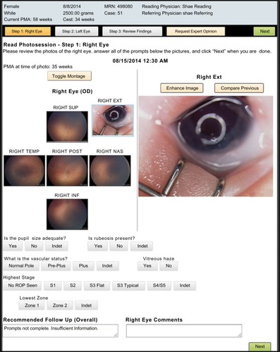
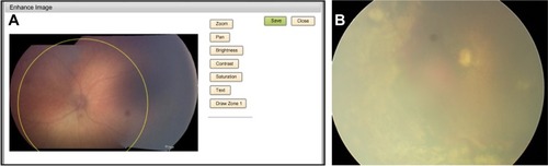
Figure 2 Computerized Zone 1 determination.
Notes: (A) Computerized software determination of Zone 1 in retinopathy of prematurity (ROP) (yellow circle). Notice the image processing capabilities (change brightness, contrast, and color saturation), which can aid in the modification of the original photo to allow a more accurate review. (B) Fundus image of the left eye of an infant with a heavily pigmented fundus obtained with a wide-field contact fundus camera (RetCam; Clarity Medical Systems, Pleasanton, CA, USA) and type 1 ROP requiring laser treatment. Media opacity is due to vitreous hemorrhage, which renders the retinal vessels difficult to discern.