Figures & data
Figure 1 Murine retinal vascular changes during OIR.
Notes: (A) Graphic representation of retinal vasculature at the microscopic level (superior image) and in microscopic cross section (inferior image) in a healthy C57Bl/6 mouse at P7 under normal conditions. Mice are normally born with avascular retinas, allowing for full observation of vascular growth postnatally. By P7, the superficial vascular plexus (ie, epiretinal vascular network) almost fully extends across the peripheral retina, and vessels begin to develop orthogonal to the retinal surface, eventually forming the intermediate and deep vascular plexus (ie, intraretinal vascular network). Full vascular maturation is usually achieved around P21–P25 in C57Bl/6 mice. (B) Depiction of vaso-obliteration during phase 1 of OIR, when mice are exposed to 75% oxygen from P7 to P12. An important distinction is that vaso-obliteration in mice occurs in the central retina, in contrast to the peripheral vaso-obliteration seen in the rat OIR model and in human ROP. During this high oxygen exposure, VEGF production is suppressed in the relatively hyperoxic central retina, resulting in vascular loss. Of note, progression of vaso-obliteration is most rapid within the first 48 hours of high oxygen exposure. (C) Upon transfer from high oxygen exposure to room air at P12, the central avascular retina experiences relative ischemia, especially given the nutritional demands of the growing retina. As a result, VEGF is produced in this ischemic zone, which activates downstream effector molecules that lead to the characteristic neovascular tufts seen in phase 2 of OIR. Neovessels primarily form at the junction of the vascular and avascular zones, with peak neovessel density reached by P17 in C57Bl/6 mice, as depicted here. (D) Depiction of the vascular repair process, where spontaneous neovessel regression coincides with waning pro-angiogenic factors and normal revascularization. This repair process is essentially complete by P25.
Abbreviations: OIR, oxygen-induced retinopathy; P, postnatal day; ROP, retinopathy of prematurity; VEGF, vascular endothelial growth factor.
Abbreviations: OIR, oxygen-induced retinopathy; P, postnatal day; ROP, retinopathy of prematurity; VEGF, vascular endothelial growth factor.
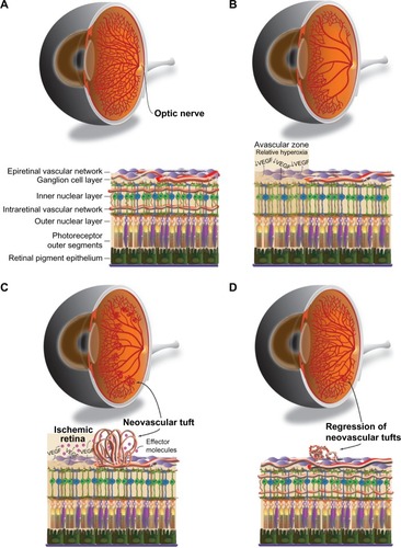
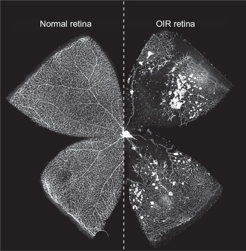
Figure 2 Control and OIR flatmounts at murine P17.
Notes: In this composite image, retinal flatmounts from C57Bl/6 mice at P17 under normal (left) and OIR (right) conditions were stained with isolectin B4 to label the vasculature (white). Normally, as seen on the left, retinal vessels branch radially from the optic disk in a uniform pattern across the entire retina. In contrast, P17 OIR mouse retinas possess extensive central vaso-obliteration with pathologic neovessels forming around the junction of the vascular and avascular zones, as evidenced by the dense, circular lectin-stained clusters seen on the right. These leaky, unorganized tufts often extend into the vitreous and reach peak density at P17, which is when neovascularization is typically assessed.
Abbreviations: OIR, oxygen-induced retinopathy; P, postnatal day.
Abbreviations: OIR, oxygen-induced retinopathy; P, postnatal day.