Figures & data
Figure 1 Schematic of retinal zones.
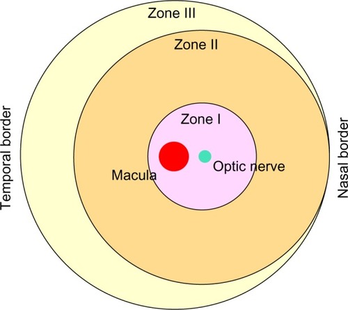
Table 1 Conventional treatments for ROP and results from clinical studies
Figure 2 Injection of ECFCs on P5 stimulates the development of the deep vascular plexus in the OIR pups prior to return to normoxic conditions on postnatal day 12.
Notes: Confocal images from flat-mounted retinas from OIR pups injected on postnatal day 5 and euthanized on postnatal day 12. The four panels show a z-stack of confocal images from retinas of OIR mouse pups on the left, and rotated (90°) images of 3-D projections of the retinas on the right showing a cross section of the retina (vitreous, left side; choroid, right side. CD34+ cells are in blue. (A) Retina from saline-injected pup, (B) from CD34+ cell-injected mouse, (C) from ECFC-injected mouse, and (D) from CD34+ cell- and ECFC-injected mouse. Blood vessels are stained with collagen IV antibody. ECFCs express GFP. ECFC incorporated into blood vessels are yellow. Scale bars, 50 μm. Original images were captured with either 10× (A, C) or 20× objective (B, C).
Abbreviations: ECFCs, endothelial colony-forming cells; OIR, oxygen-induced retinopathy; SVP, superficial vascular plexus; DVP, deep vascular plexus.
Abbreviations: ECFCs, endothelial colony-forming cells; OIR, oxygen-induced retinopathy; SVP, superficial vascular plexus; DVP, deep vascular plexus.
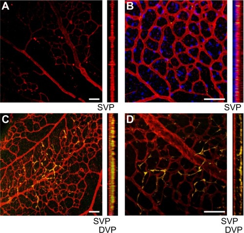
Figure 3 Ischemic injury on retinal micro-blood vessel results in loss of pericytes and endothelial cells and vaso-occlusion.
Notes: MSCs and ASCs differentiate into pericytes. Cell-surface receptors on intravitreally injected iPS-ECFCs or ECFCs interact with paracrine-released factors from CD34+ cells to differentiate into endothelial cells.
Abbreviations: MSCs, mesenchymal stem cells; RBC, red blood cells; ASCs, adipose-derived stem cells; iPS-ECFCs, induced pluripotent stem endothelial colony-forming cells.
Abbreviations: MSCs, mesenchymal stem cells; RBC, red blood cells; ASCs, adipose-derived stem cells; iPS-ECFCs, induced pluripotent stem endothelial colony-forming cells.
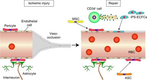
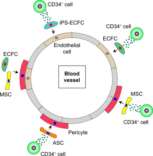
Figure 4 Combination of various cell types that repair damage to ischemia-derived injuries.
Abbreviations: iPS-ECFC, induced pluripotent stem endothelial colony-forming cell; MSC, mesenchymal stem cell; ASC, adipose-derived stem cell; ECFC, endothelial colony-forming cells.