Figures & data
Figure 1 Example of severe dysgenesis with unregressed stalk tissue and primitive retinal development.
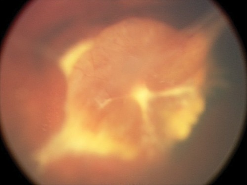
Figure 2 Fluorescein angiogram of a Wnt-associated vitreoretinopathy patient with an Fzd4 mutation.
Note: The avascular periphery, tractional fold, and anomalous capillary formation is noted.
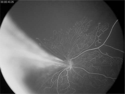
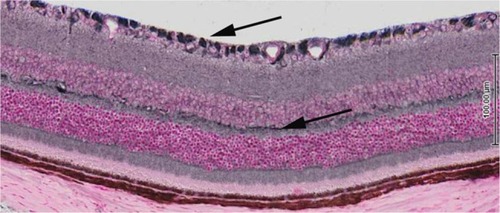
Figure 3 Cross-section of mouse retina post-natal day 21 demonstrating expression of the LGR4 receptor throughout the retina.
Note: Arrows depict higher expression in the retinal ganglion cells and outer plexiform layer.