Figures & data
Table 1 Reports of refractive error after the use of anti-vascular endothelial growth factor agents for the treatment of retinopathy of prematurity
Figure 1 Zone I distribution of refractive error by treatment modality.
Notes: Distribution of spherical equivalent refractive error at age 2.5 years in eyes that received treatment for stage 3+ retinopathy of prematurity or aggressive posterior retinopathy of prematurity in the Bevacizumab Eliminates the Angiogenic Threat for Retinopathy of Prematurity clinical trial.Citation1 Data presented according to treatment modality: red, laser without recurrence; brown, laser with recurrence; light blue, intravitreal bevacizumab without recurrence; dark blue, intravitreal bevacizumab with recurrence. Reproduced with permission from JAMA Ophthalmol. 2014;132(11):1327–1333. Copyright ©2014 American Medical Association. All rights reserved.Citation1
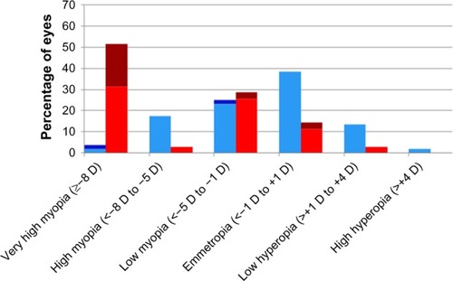
Figure 2 Zone II posterior distribution of refractive error by treatment modality.
Notes: Distribution of spherical equivalent refractive error at age 2.5 years in eyes that received treatment for stage 3+ retinopathy of prematurity or aggressive posterior retinopathy of prematurity in the Bevacizumab Eliminates the Angiogenic Threat for Retinopathy of Prematurity clinical trial.Citation1 Data presented according to treatment modality: red, laser without recurrence; brown, laser with recurrence; light blue, intravitreal bevacizumab without recurrence; dark blue, intravitreal bevacizumab with recurrence. Reproduced with permission from JAMA Ophthalmol. 2014;132(11):1327–1333. Copyright © 2014 American Medical Association. All rights reserved.Citation1
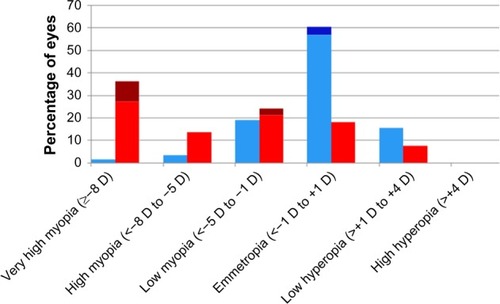
Table 2 Cycloplegic retinoscopic refractive error at age 2.5 years in the Bevacizumab Eliminates the Angiogenic Threat for Retinopathy of Prematurity clinical trialCitation1
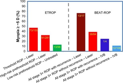
Figure 3 Percentage of eyes ≥−5 D in ETROP and BEAT-ROP by ROP severity at treatment.
Notes: Percentage of eyes with myopia ≥−5 D in the ETROP and BEAT-ROP clinical trials at ages 3 and 2.5 years, respectively. Data presented according to ROP severity at treatment (threshold, high-risk prethreshold, low-risk prethreshold, and all stage 3+) by treatment modality utilized (laser and IVB) in the respective clinical trials. Data from ETROP,Citation3 and BEAT-ROP.Citation1
Abbreviations: ROP, retinopathy of prematurity; IVB, intravitreal bevacizumab.
Abbreviations: ROP, retinopathy of prematurity; IVB, intravitreal bevacizumab.