
Figures & data
Figure 1 Intraoral appearance of the patient in Case 1 (A–E) There were “cottage cheese-like” lesions involving the entire oral cavity on the first visit (F–J) After two weeks, the lesions healed.
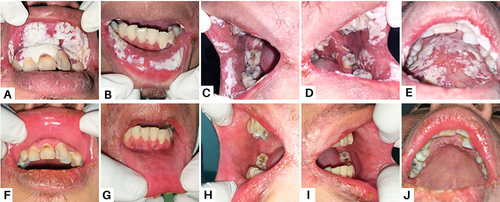
Table 1 Serological Examination Result
Figure 2 Clinical features of the patient in Case 2 (A–E) On the first visit, white spots on the dorsal tongue and erythematous areas (F–J) The lesions have improved after one month.
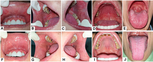
Figure 3 Clinical presentation of the patient Case 3 (A and B) On the first visit, the thrush was present as yellowish-white plaques on the tongue (C) A single ulcer on the buccal mucosa (D and E) A painful, primary, single ulcer on the oropharynx (F–J) The lesions had entirely resolved after four weeks.
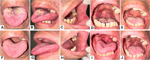
Figure 4 Intraoral lesions in patient Case 4 (A, B, D and E) On the first visit, there were extensive white plaques on the palate and tongue (C) A painful single ulcer with an irregular, yellowish base on the right lateral tongue (F–J) After one month, the lesions recovered.
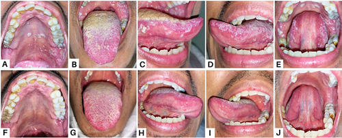
Figure 5 Intraoral condition in patient Case 5 (A–D) On the first visit, white plaques resembling curdled milk were present on the labial mucosa and buccal mucosa (E) Diffuse erythema of the hard palate (F–J) After four weeks, the lesions disappeared.
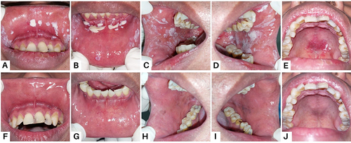
Table 2 A Brief Comparison of Five Cases