Figures & data
Figure 1 Pathophysiology of nonalcoholic fatty liver disease (NAFLD).
Notes: 1. Adipocyte mobilization increases serum concentration of FFA and de novo synthesis of triglyceride. 2. Increased FFA oxidation in the mitochondria exceeds the capacity of the mitochondria, causing oxidative stress, which impairs insulin sensitivity and autophagy activation to digest defective proteins. 3. Oxidative stress promotes phosphorylation and degradation of mesenchymal IKKB, accompanied by activated NF-κB translocation into the nucleus, causing inflammatory cascades and liver injury. 4. Obesity causes ER stress, which causes insulin resistance and stimulates autophagy due to an increase in unfolded proteins. Defective autophagy aggravates ER stress. 5. Short-term insulin resistance activates autophagy while long-term lipotoxicity impairs autophagy. 6. Energy produced by autophagy and oxidation fuels HSC activation, which promotes hepatic fibrosis. 7. NAFLD develops from steatosis, steatohepatitis, fibrosis, and hepatoma.
Abbreviations: FFA, free fatty acid; ER, endoplasmic reticulum; HSC, hepatic stellate cells.
Abbreviations: FFA, free fatty acid; ER, endoplasmic reticulum; HSC, hepatic stellate cells.
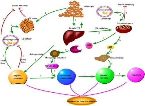
Figure 2 Electron micrograph showing ultrastructure of hepatocytes from a NAFLD mouse model.
Notes: Black arrow indicates autophagic vacuoles with a double-membrane structure that enclose lipid droplets and other defective organelles. White arrow shows autolysosome digesting contents. Original magnification: 100,000×.
Abbreviation: NAFLD, nonalcoholic fatty liver disease.
Abbreviation: NAFLD, nonalcoholic fatty liver disease.
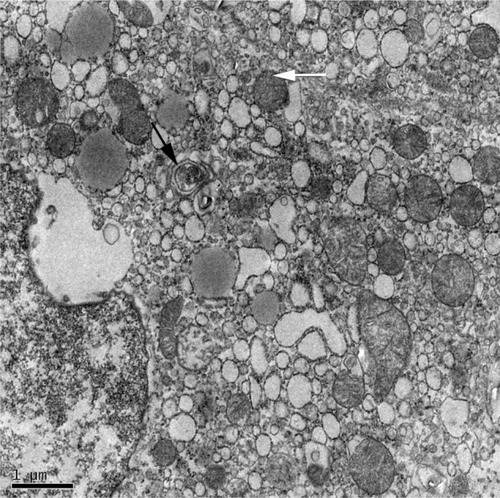
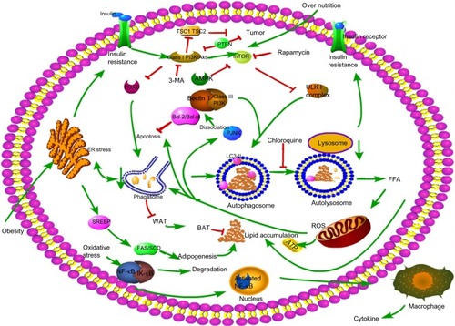
Figure 3 Molecular mechanisms involved in autophagy and NAFLD.
Notes: 1. A small vascular sac called an isolation membrane is formed to enclose lipid droplets, which results in a double membrane structure called an autophagosome, which subsequently fuses with a lysosome to degrade lipid droplets into FFA. 2. Growth factor activates the PI3K/Akt signaling pathway to active mTOR, leading to phosphorylation of the ULK1 complex, resulting in inhibition of autophagy formation. Short-term resistance may activate autophagy via inhibition of mTOR, while long-term lipotoxicity impairs autophagy by inhibition of the FOXO pathway. 3. The class III PI3K/Beclin-1 complex positively regulates autophagy. 4. Increased FFA oxidation causes mitochondrial oxidative stress and activates autophagy. ROS also promotes cell apoptosis and JNK phosphorylation, which stimulates Bcl-2 phosphorylation and Bcl-2/Beclin-1 disassociation, resulting in apoptosis and autophagy, respectively. Likewise, ROS promotes NF-κB translocation into the nucleus, causing inflammatory cascades and liver injury. 5. Obesity causes ER stress, which activates autophagy and promotes adipogenesis-related gene expression such as FAS, SCD, leading to the synthesis of adipocytes. 6. Autophagy may show an inhibitory effect on adipocyte differentiation from WAT to BAT. 7. MP-activated protein kinase (AMPK) inhibits mTOR, and ultimately activates autophagy.
Abbreviations: NAFLD, nonalcoholic fatty liver disease; FFA, free fatty acid; ROS, reactive oxygen species; ER, endoplasmic reticulum; WAT, white adipose tissue; BAT, brown adipose tissue.
Abbreviations: NAFLD, nonalcoholic fatty liver disease; FFA, free fatty acid; ROS, reactive oxygen species; ER, endoplasmic reticulum; WAT, white adipose tissue; BAT, brown adipose tissue.