Figures & data
Figure 1 Etomoxir combination improves radiation efficacy against sphere formation by H460 lung epithelial cancer cells.
Notes: (A) Experimental scheme. (B) At the end of experiment (day 6), sphere images were captured and representative images are presented (100×). (C) Average sphere area presented as mean±SEM. (D) Number of spheres in each group presented as mean±SEM. (E) Experimental scheme. (F) At the end of 7 days, number of colonies with ≥50 cells were counted and presented as mean±SEM in the bar diagram. In the bar diagram, a represents control group, b represents etomoxir alone, c represents radiation alone, and d represents radiation plus etomoxir.*p<0.001; $p<0.01; #p<0.05.
Abbreviations: Eto, etomoxir; Rad, radiation; SEM, standard error of the mean.
Abbreviations: Eto, etomoxir; Rad, radiation; SEM, standard error of the mean.
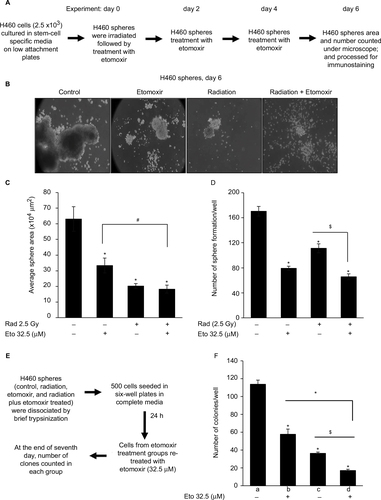
Figure 2 Etomoxir treatment following radiation exposure reduces hypoxic areas in H460 spheres.
Notes: H460 spheres were irradiated (2.5 Gy) and then treated with etomoxir 32.5 µM. Spheres were re-treated with etomoxir on experiment day 2 and 4. At the end of day 6, H460 spheres were treated with pimonidazole (200 µM) for 2 h and then processed for immunofluorescence. Representative confocal images are shown (at 200× magnification). The bar diagram represents the mean fluorescence intensity per unit sphere area as mean±SEM. *p<0.001; $p<0.01.
Abbreviations: DAPI, 4′,6-diamidino-2-phenylindole; Eto, etomoxir; Rad, radiation; SEM, standard error of the mean.
Abbreviations: DAPI, 4′,6-diamidino-2-phenylindole; Eto, etomoxir; Rad, radiation; SEM, standard error of the mean.
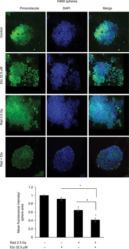
Figure 3 Etomoxir treatment following radiation exposure reduces the expression of proliferation and stemness biomarkers in H460 spheres.
Notes: (A–C) 5 × 10³ H460 cells were plated in six-well ultralow attachment plates for sphere formation for 6 days. Thereafter, spheres were irradiated with a dose of 2.5 Gy and then treated with etomoxir 32.5 µM. Spheres were retreated with etomoxir on experiment days 2 and 4. At the end of day 6 after first etomoxir treatment, H460 spheres were processed to analyze (A) Ki-67, (B) cyclin D1, and (C) CD44 expression by immunofluorescence as described in the “Methods” section. Representative images are shown (at 200× magnification). (D) The bar diagram represents the mean fluorescence intensity per unit sphere area for Ki-67, cyclin D1, and CD44 as mean±SEM. *p<0.001; $p<0.01; #p<0.05.
Abbreviations: DAPI, 4′,6-diamidino-2-phenylindole; Eto, etomoxir; Rad, radiation; SEM, standard error of the mean.
Abbreviations: DAPI, 4′,6-diamidino-2-phenylindole; Eto, etomoxir; Rad, radiation; SEM, standard error of the mean.
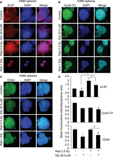
Figure 4 Etomoxir treatment following radiation exposure reduces CPT1 expression in H460 spheres.
Notes: 5 × 10³ H460 cells were plated in six-well ultralow attachment plates for sphere formation for 6 days. Thereafter, spheres were irradiated with a dose of 2.5 Gy and then treated with etomoxir 32.5 µM. Spheres were retreated with etomoxir on experiment days 2 and 4. At the end of day 6 after first etomoxir treatment, H460 spheres were processed to analyze CPT1A expression by immunofluorescence as described in the “Methods” section. Representative images are shown (at 200× magnification). The bar diagram represents the mean fluorescence intensity per unit sphere area as mean±SEM. *p<0.001; $p<0.01.
Abbreviations: CPT1A, carnitine palmitoyltransferase 1A; DAPI, 4′,6-diamidino-2-phenylindole; Eto, etomoxir; Rad, radiation; SEM, standard error of the mean.
Abbreviations: CPT1A, carnitine palmitoyltransferase 1A; DAPI, 4′,6-diamidino-2-phenylindole; Eto, etomoxir; Rad, radiation; SEM, standard error of the mean.
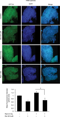
Figure 5 Radiation treatment effectively reduces sphere size and hypoxic areas in CPT1A knockdown LNCaP cells.
Notes: LNCaP spheres were irradiated (2.5 Gy) and then treated with etomoxir 32.5 µM. At the end of day 6, LNCaP spheres number was counted and presented as mean±SEM in the bar diagram (A). Thereafter, spheres in each group were dissociated into single cells and plated 1,000 cells/well in ultralow attachment six-well plates. Cells were treated with etomoxir 32.5 µM after 24 h of seeding and every 48 h thereafter. Number of spheres were counted after day 6 of treatment and presented as mean±SEM in the bar diagram (B). In the bar diagram, a represents control group, b represents etomoxir alone, c represents radiation alone, and d represents radiation plus etomoxir. (C, D) Vector control LNCaP cells and LNCaP CPT1A KD cells were cultured to form spheres and then treated with radiation (2.5 Gy). After 6 days, sphere number and area were measured and presented as mean±SEM in the bar diagrams. (E) LNCaP CPT1 KD spheres were treated with 200 µM of pimonidazole for 2 h and processed for immunofluorescence. Representative confocal images are shown (at 200× magnification). The bar diagram represents the mean fluorescence intensity per unit sphere area as mean±SEM. *p<0.001; $p<0.01; #p<0.05.
Abbreviations: CPT1A, carnitine palmitoyltransferase IA; DAPI, 4′,6-diamidino-2-phenylindole; Eto, etomoxir; KD, knockdown; Rad, radiation; SEM, standard error of the mean.
Abbreviations: CPT1A, carnitine palmitoyltransferase IA; DAPI, 4′,6-diamidino-2-phenylindole; Eto, etomoxir; KD, knockdown; Rad, radiation; SEM, standard error of the mean.
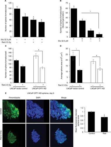
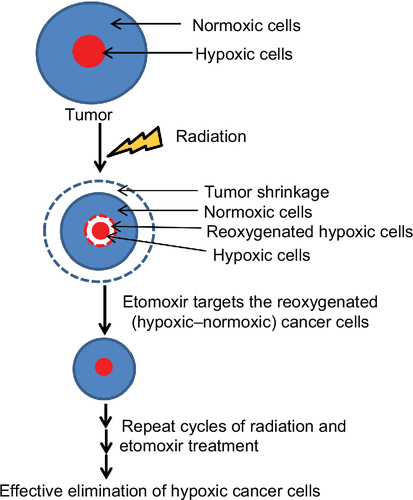
Figure 6 Proposed hypothesis.
Note: Etomoxir, an inhibitor of β-oxidation, treatment following radiation exposure could effectively eliminate hypoxic cells in the tumor, inhibiting tumor growth and preventing disease relapse.