
Figures & data
Figure 1 Pathogenesis of invasive dermatophytosis.
Notes: Physical trauma impairs the epidermal barrier. Penetration of the dermatophytes into the skin causes a granulomatous, inflammatory response, including neutrophils (N), eosinophils (E), lymphocytes (T), macrophages (M), and multinuclear giant cells (MGC). Majocchi’s granuloma (A), mycetoma (B), deeper dermatophytosis (C), and disseminated dermatophytosis (D).
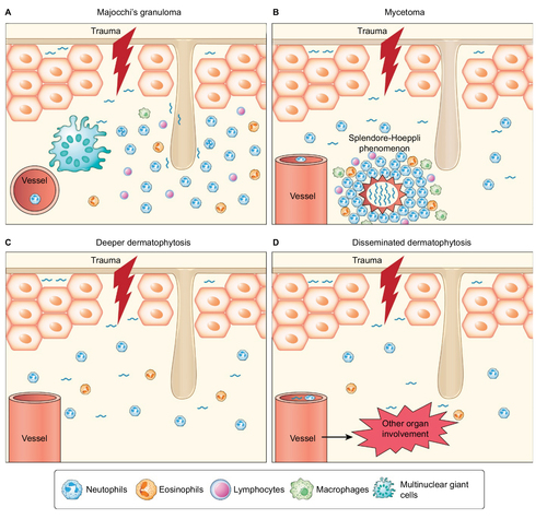
Figure 2 Multiple erythematous papules and nodules with scales and/or crusts are located on the anterior surface of the abdomen in a patient with Majocchi’s granuloma (A). Erythematous plaque with pustules, scales, and crusts on the lateral side of the arm in a patient with Majocchi’s granuloma (B).
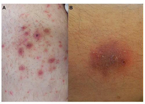
Figure 3 Clinical characteristics of the patients with MG reported in the literature: location of the lesions, sex, immunity, predisposing factors, and type of lesion.
Abbreviation: MG, Majocchi’s granuloma.
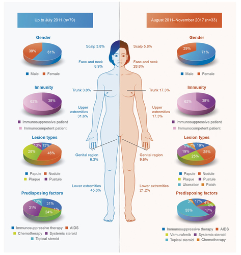
Figure 4 Histopathological findings of a patient with MG.
Notes: (A) The histopathology showed perifollicular, granulomatous inflammation (arrows). (B) Hyphae (arrows) are seen with great magnification. (C) Perifollicular spores (arrows) were positively stained with PAS staining. (D) In the HE-stained slides, spores (arrows) showed autofluorescence under an immunofluorescence microscope. (A, HE ×100; B, D, HE ×1000; C, PAS ×1000).
Abbreviations: MG, Majocchi’s granuloma; PAS, periodic acid-Schiff staining; HE, hematoxylin and eosin.
Abbreviations: MG, Majocchi’s granuloma; PAS, periodic acid-Schiff staining; HE, hematoxylin and eosin.
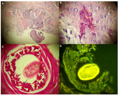
Table 1 The causative fungi that were isolated from patients with MG