Figures & data
Table 1 Epidemiologic and clinical characteristics of five patients with recurrent (M) XDR-TB following drug-susceptible TB
Table 2 HIV drug resistance profiles at TRUTH study entry and exit visits
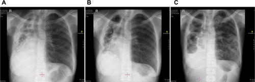
Figure S1 Chest X-rays of Patient 1 depicting (A) infiltrates on both lungs; (B) persistent fibrosis in the left lung; and (C) cavities in the left lung.
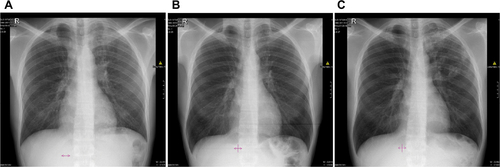
Figure S2 Chest X-rays of Patient 2 depicting (A) infiltrates in both lungs and cavities in the left lung; (B) right upper lobe fibrosis; and (C) infiltrates in the right lung and infiltrates and new lesions in the left lung.
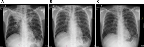
Figure S3 Chest X-rays of Patient 3 depicting (A) cavities and infiltrates in both lungs; (B) cavities and infiltrates on the right lung and fibrosis in the left lung; and (C) cavities, infiltrates, and patchy consolidation in the right lung and consolidation in mid and lower zones of the left lung.
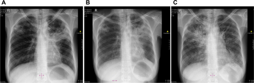
Figure S4 Chest X-rays of Patient 4 depicting (A) vast damage to the right lung with cavities, infiltrates, pleural disease, shrinkage, and destruction. Minimal damage to the left lung; (B) residual widespread damage to the right lung; and (C) right lung bullae and new pleural effusion. New infiltrates in left lung.