
Figures & data
Figure 1 Functional domain and conservation of rpsL (A) and gyrA (B) mutation sites in Mycobacterium spp.
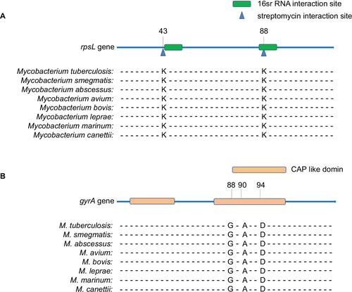
Table 1 Mutation types of single- and dual-resistant Mycobacterium smegmatis mutants under the selection of antibiotics
Figure 2 Relative fitness of Mycobacterium smegmatis mutants.
Notes: (A) Relative fitness of STR-resistant M. smegmatis. (B) Relative fitness of FLQ-resistant M. smegmatis. (C) Relative fitness of STR and FLQ dual-resistant M. smegmatis. Gray bar is for the STR-resistant M. smegmatis, turquoise is for FLQ-resistant, and purple is for the STR–FLQ dual-resistant M. smegmatis.
Abbreviations: FLQ, fluoroquinolones; STR, streptomycin.
Abbreviations: FLQ, fluoroquinolones; STR, streptomycin.
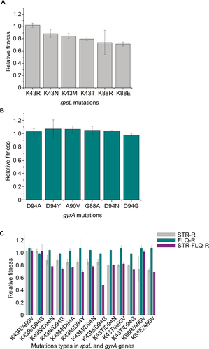
Figure 3 Frequency of dual-mutation of rpsL and gyrA found in clinical Mycobacterium tuberculosis isolates.
Notes: (A) Mutation combinations of clinical M. tuberculosis isolates from Sichuan. (B) Mutation combinations of 3,056 M. tuberculosis genome sequence in database.
Abbreviation: MTB, Mycobacterium tuberculosis.
Abbreviation: MTB, Mycobacterium tuberculosis.
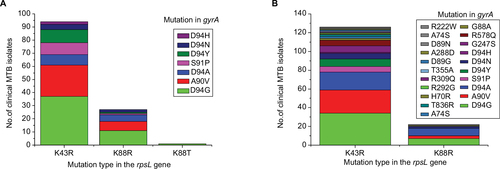
Table 2 Gene mutations in clinical STR- and FLQ-resistant Mycobacterium tuberculosis isolates
Figure S1 Model of mutations appearing in rpsL and gyrA in this study.
Notes: (A) Structure of Escherichia coli rpsL (Protein Data Bank accession number 5U4I). The position of K43 residue (yellow) and K88 residue (red) screened in this study was highly conserved between E. coli and Mycobacterium tuberculosis. (B) M. tuberculosis gyrA (Protein Data Bank accession number 3IFZ). The position of G88 residue (yellow), A90 residue (red), and D94 residue (cyan) screened in present study is shown. Amino acids were placed in the cartoon structure in dots format using the PyMOL Viewer.
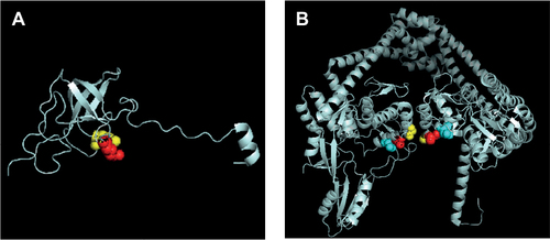
Figure S2 The alignment of rpsL in M. tuberculosis and E. coli.
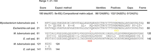
Table S1 Epistasis (ε) in mutants resistant to STR and FLQ