Figures & data
Table 1 Clinical data of the patients with tuberculosis of the parotid nodes
Figure 1 The multiple tuberculous lesions of right parotid nodes are linearly arranged on sagittal CT image.
Notes: Tuberculosis of the left parotid lymph node in a 28-year-old female. (A) Volume rendering image (white arrow); (B) local rupture of parotid fascia and thickened adjacent skin (white arrow). Axial and coronal images (C, D) show an irregular cyst-like enhanced mass (white arrow), located in the superficial lobe and close to the surface of the sternocleidomastoid which involves parotid fascia; the neighboring skin is thickened and adhered (C, D) (white arrowhead).
Abbreviation: CT, computed tomography.
Abbreviation: CT, computed tomography.
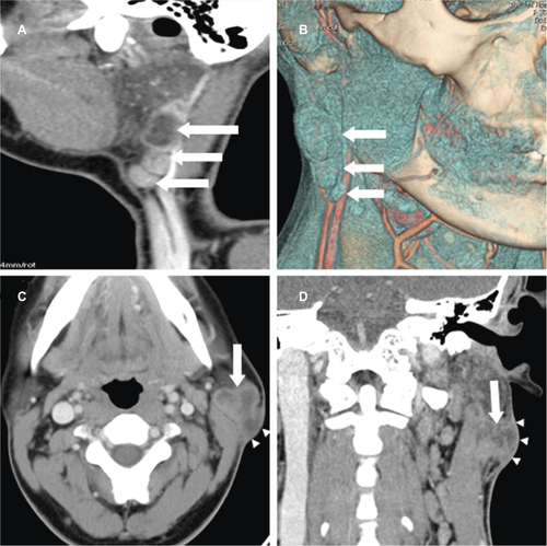
Figure 2 Homogenous enhancement.
Notes: Tuberculosis of the right parotid node in a 30-year-old female. Axial CT image (A) shows a regular mass (white arrow) with homogeneous enhancement, which is located in the superficial lobe, accompanied by cervical lymphadenopathy level IIa and level IIb (black arrow). Thick-walled circular enhancement. Tuberculosis of the right parotid nodes in a 37-year-old female. Axial CT image (B) shows a smooth rounded nodule (white arrow) with thick-walled circular enhancement and well-defined border, which is located in the superficial lobe. Garland-like enhancement. Tuberculosis of the right parotid lymph nodes in a 37-year-old female. Axial CT image (C) shows a lobulated nodule (white arrow) with garland-like enhancement, which is located in the superficial lobe. Irregular cyst-like enhancement. Tuberculosis of the left parotid lymph nodes in a 16-year-old female. Axial CT image (D) shows a mass (white arrow) with irregular cyst-like enhancement, which is located in the superficial lobe.
Abbreviation: CT, computed tomography.
Abbreviation: CT, computed tomography.
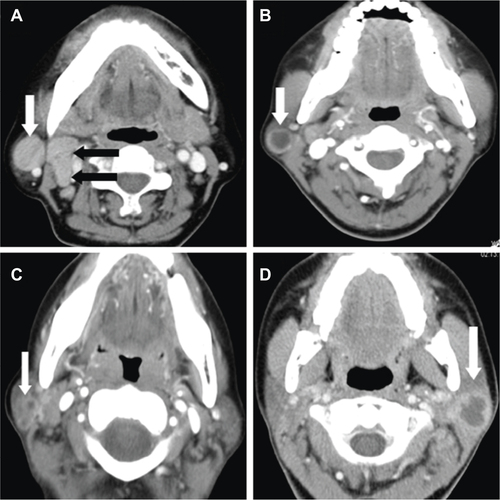
Table 2 Distribution of cervical lymphadenopathy (n=25)
Table 3 Magnetic resonance findings of patients with TB of the parotid nodes
Figure 3 Tuberculosis of the left parotid lymph node in a 19-year-old male.
Notes: The lesion is located in the superficial lobe, with isointensity on axial T1WI (A) (black arrow), slight hyperintensity on coronal T2WI-FS (B) (white arrow), and obvious enhancement (C, D). The surrounding parotid parenchymal edema (black arrowhead) and thickened adjacent fascia (white arrowhead) are shown clearly on coronal T2WI-FS (B) and obvious enhancement is shown on post-contrast images (C, D).
Abbreviations: T1WI, T1-weighted image; T2WI-FS, T2-weighted image with fat saturation.
Abbreviations: T1WI, T1-weighted image; T2WI-FS, T2-weighted image with fat saturation.
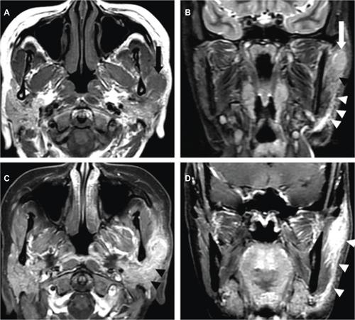
Figure 4 Tuberculosis of the right parotid lymph node in a 16-year-old male.
Notes: Coronal T1WI (A) shows a nodule with isointense signal intensity (black arrow), which is located in the superficial lobe. The lesion also shows isointense signal intensity (black arrow) on coronal T2WI (B). Bilateral cervical lymphadenopathy is shown on coronal T1WI (C, D) (white arrow). The thickened parotid fascia is shown clearly on coronal T1WI and T2WI (A, B) (white arrowhead).
Abbreviations: T1WI, T1-weighted image; T2WI, T2-weighted image.
Abbreviations: T1WI, T1-weighted image; T2WI, T2-weighted image.
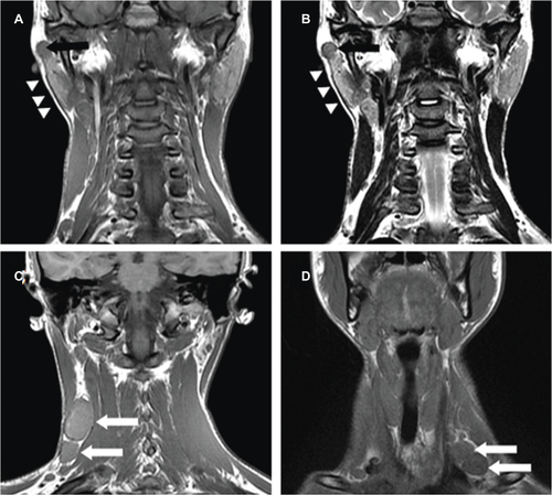
Figure 5 Tuberculosis of the right parotid lymph node in a 58-year-old female.
Notes: The lesion is located in the superficial lobe with isointense signal intensity on axial T1WI (A) (white arrow) and heperintense signal intensity on axial T2WI-FS images (B) (black arrow). Markedly hyperintense signal intensity is shown on DWI (C) (white arrow) and hypointense signal intensity on ADC map (D) (black arrow).
Abbreviations: T1WI, T1-weighted image; T2WI-FS, T2-weighted image with fat saturation; DWI, diffusion-weighted imaging; ADC, apparent diffusion coefficient.
Abbreviations: T1WI, T1-weighted image; T2WI-FS, T2-weighted image with fat saturation; DWI, diffusion-weighted imaging; ADC, apparent diffusion coefficient.
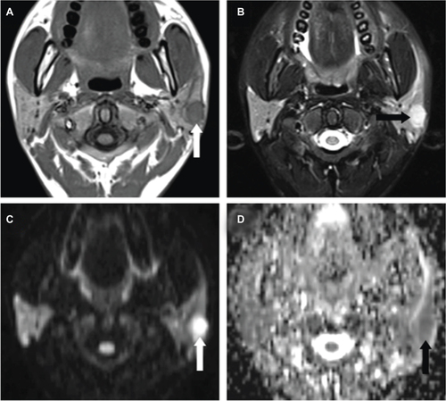
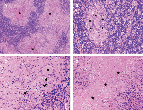
Figure 6 Histopathology of parotid nodes.
Notes: Tuberculous nodules (A) (black star) with typical Langhans giant cell (B, C) (black star), epithelioid macrophages (C) (black arrowhead), and caseous necrosis (D) (black star). (A: H&E stain, magnification ×100; B, C: H&E stain, magnification ×400; D: H&E stain, magnification ×200).