Figures & data
Figure 1 Identification of E. coli with or without lysis buffer.
Notes: Bacilli were found with (A) and without (B) lysis buffer. The bioMérieux blood culture system was used in both conditions. Arrows indicate bacilli.
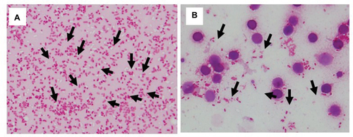
Figure 2 Display within the BioTyper 2.0 graphic view in the case of E. coli.
Notes: E. coli with (A) or without (B) lysis buffer, respectively. The mass spectra show the difference in peaks (presence or absence) and their intensities between the sample spectrum and those of bacteria identified at first pass by BioTyper. The upper part of the figure within the inner windows shows the unknown spectrum containing perfectly matching peaks (0–200 ppm) in green, imperfectly matching peaks (200–500 ppm) in yellow, and nonmatching peaks in red. The lower part (blue) shows the dedicated main spectrum included in the database. MALDI-TOF MS score values were 2.315 (A) and 1.7799 (B), respectively. The bioMérieux blood culture system was used in both conditions.
Abbreviation: MALDI-TOF MS, matrix-assisted laser desorption/ionization time-of-flight mass spectrometry.
Abbreviation: MALDI-TOF MS, matrix-assisted laser desorption/ionization time-of-flight mass spectrometry.
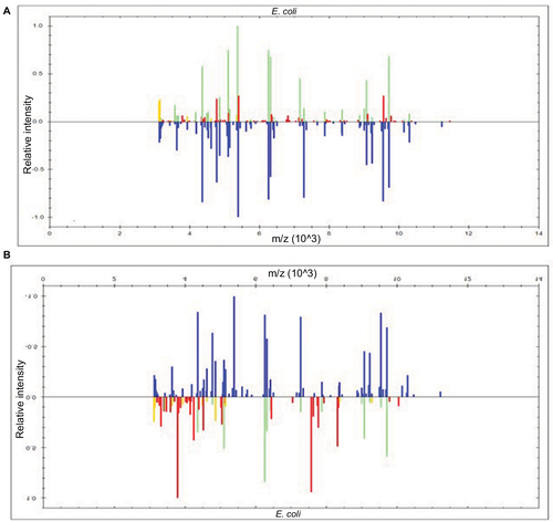
Table 1 Detection efficiency of bacteria from blood culture broth by TOF-MS with or without lysis buffer
Figure 3 Identification of S. pneumoniae with or without lysis buffer.
Notes: (A, B, and C) Lysis buffer was used, (D, E, and F) lysis buffer was not used. (A) and (B) BD, (C) and (D) bioMérieux, (E) and (F) Oxoid blood culture systems. Arrows indicate bacilli.
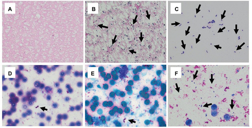
Figure 4 Identification of H. influenzae with or without lysis buffer.
Notes: (A, B, and C) Lysis buffer was used, (D, E, and F) lysis buffer was not used. (A) and (D) BD, (C) and (D) bioMérieux, (E) and (F) Oxoid blood culture systems. Arrows indicate bacilli.
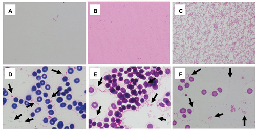
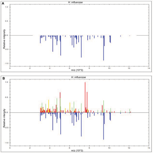
Figure 5 Display within the BioTyper 2.0 graphic view in the case of H. influenzae.
Notes: H. influenzae with (A) or without (B) lysis buffer, respectively. The mass spectra show the difference in peaks (presence or absence) and their intensities between the sample spectrum and those of bacteria identified at first pass by BioTyper. The upper part of the figure within the inner windows shows the unknown spectrum containing perfectly matching peaks (0–200 ppm) in green, imperfectly matching peaks (200–500 ppm) in yellow, and nonmatching peaks in red. The lower part (blue) shows the dedicated main spectrum included in the database. MALDI-TOF MS score values were none (A) and 1.921 (B), respectively. The bioMérieux blood culture system was used in both conditions.
Abbreviation: MALDI-TOF MS, matrix-assisted laser desorption/ionization time-of-flight mass spectrometry.
Abbreviation: MALDI-TOF MS, matrix-assisted laser desorption/ionization time-of-flight mass spectrometry.