Figures & data
Table 1 Bacterial strains used in decontamination assays
Figure 1 Phage activity against S. aureus, E. coli, and P. aeruginosa (ATCC or MDR strains). Bacteriophage activity was verified by spot tests. Briefly, after suspension in soft agar, bacterial cultures were overlaid on TSA plates; serially diluted phage stocks were added to bacterial lawns, checking their lytic activity after 24 hours of incubation at 37°C. Results are representative of triplicate samples.
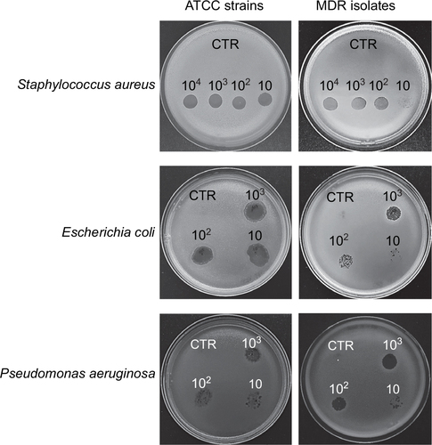
Figure 2 Reduction of bacterial load on different types of hard surfaces treated with specific phages. One hundred CFU of each ATCC bacterial strain (S. aureus, E. coli, and P. aeruginosa) were spread on the indicated sterile surfaces, allowed to dry, and subsequently treated with the specific phages, suspended in PBS, at the indicated MOI. After 1, 3, 6, and 24 hours at room temperature, residual viable cells were measured by Rodac sampling with specific selective media and subsequent CFU count after 24 hours of incubation at 37°C. Negative CTR(−) samples were treated with PBS alone. Positive CTR(+) samples were treated with denatured alcohol. Results represent the mean ± SD of triplicate samples in two independent assays per bacterial target.
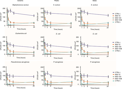
Figure 3 Reduction of MDR hospital isolates on hard surfaces treated with specific phages. One hundred CFU of each bacterial isolate (wild-type S. aureus, E. coli, and P. aeruginosa strains) were spread on ceramic, plastic, or glass surfaces, allowed to dry, and subsequently treated with the specific phages, suspended in PBS, at 1000 MOI. After 1, 3, 6, and 24 hours at room temperature, residual viable cells were measured by Rodac sampling with specific selective media, and subsequent CFU count after 24 hours of incubation at 37°C. Negative CTR(−) samples were treated with PBS alone. Positive CTR(+) samples were treated with denatured alcohol. Results represent the mean of triplicate samples in two independent experiments, for each surface type. As no significant differences were observed between surface types, graphed values represent the mean ± SD of all the measured samples (18 total samples).
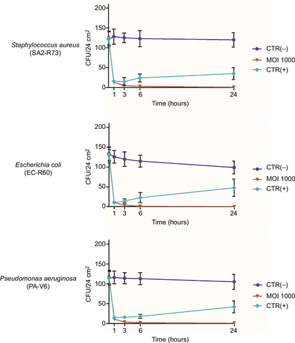
Figure 4 Phage stability in PCHS detergent. Phage stocks were suspended in PBS or in PCHS detergent diluted in water as indicated by the manufacturer, and kept at room temperature in closed plastic tubes for 1, 2, 3, or 7 days. After the indicated times, aliquots were collected and titrated by PFU counting on the corresponding ATCC bacterial target. Samples were performed in duplicate. Pictures refer to anti-E. coli phages. Superimposable results were obtained with the phages directed against S. aureus and P. aeruginosa.

Table 2 PFU titration of phage preparation in detergent and PBS
Figure 5 Reduction of Staphylococcus aureus contamination in situ, by a combined phage–probiotic detergent. About 100 CFU of S. aureus (ATCC strain) per 24 cm2 were uniformly spread on the surface of a ceramic sink. After 24 hours, the artificially contaminated surface was treated with water (CTR), probiotic detergent alone (PCHS), anti-staphylococcal phages in PBS alone (phages), or probiotic detergent including anti-staphylococcal phages (PCHS + phages). Phages were used at 1000 MOI. After 1 hour, and 1, 3, and 15 days, surfaces were sampled by Baird–Parker Rodac plates, and residual S. aureus viable cells were counted by enumerating black round colonies after 24 hours of incubation at 37°C. PCHS-Bacilli gave rise to gray-brown irregular colonies on Baird–Parker medium, easily distinguishable from the S. aureus ones. Results are representative of duplicate samples in three independent experiments.
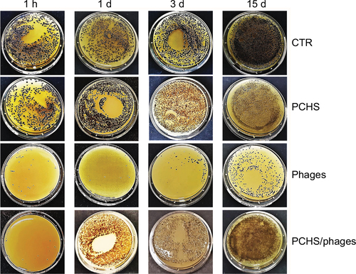
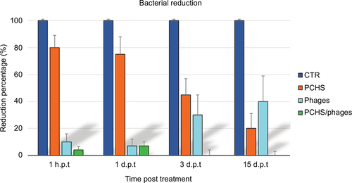
Figure 6 Average reduction of Staphylococcus aureus contamination in situ, by a combined phage–probiotic detergent. Surfaces were artificially contaminated with S. aureus and subsequently treated as described in Figure 5. Results are expressed as mean value ± SD of duplicate samples from three independent experiments. CTR, water; PCHS, probiotic detergent alone; phages, anti-staphylococcal phages in PBS alone; PCHS + phages, probiotic detergent including anti-staphylococcal phages.