
Figures & data
Figure 1 The Erta-NS prevalence rates (%) among the isolates of three leading species (Escherichia coli, Klebsiella pneumoniae, and various Enterobacter species; black bars) causing IAI, and the corresponding prevalence rates of CPE (%; gray bars) on the Erta-NS-IAI isolates in the Asia-Pacific countries (or regions) participating in this IAI study, from 2008 through 2014.
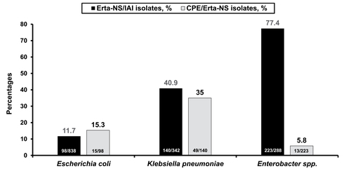
Figure 2 The prevalence rates (%) of Erta-NS isolates among the isolates causing IAI validated by testing for presence of the gene(s) encoding β-lactamase(s) (black bars) and the prevalence rates of carbapenemase producing CPE (n>10) among the Erta-NS isolates (gray bars; only isolate numbers >10 per country or region during the study period are included herein) in the Asia-Pacific countries (or regions) participating in this IAI study, from 2008 through 2014.
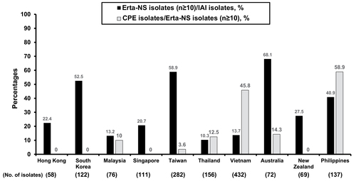
Figure 3 The prevalence rates (%) of Erta-NS isolates among the isolates that caused IAI (n=1,564) and were positive for harboring gene(s) encoding β-lactamase(s) (black bars), and the prevalence rates of carbapenemase producing CPE among the Erta-NS isolates (gray bars) in the various age populations of patients hospitalized in the Asia-Pacific countries (or regions) participating in this IAI study, from 2008 through 2014.
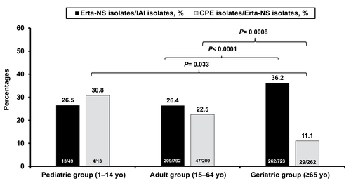
Figure 4 The prevalence rates of Erta-NS isolates among those causing IAI (n=1,564) and those that were confirmed to possess gene(s) encoding β-lactamase(s) (black bars), and the percentages of carbapenemase producing CPE among the Erta-NS isolates (gray bars) in the ICU and various ward settings in the Asia-Pacific countries (or regions) participating in this IAI study, from 2008 through 2014.
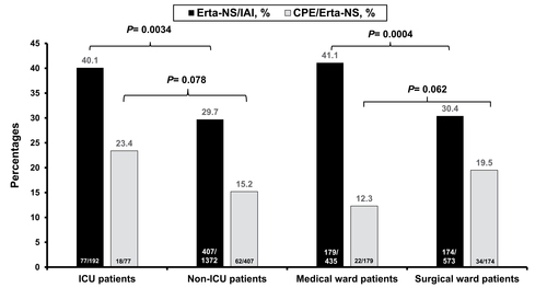
Table 1 MICs against imipenem, and the imipenem MIC50, MIC90 levels of diverse carbapenemases on the ertapenem-non-susceptible Enterobacteriaceae isolates causing intra-abdominal infections among patients hospitalized in the Asia-Pacific region from 2008 through 2014
Table 2 Comparison of the variables of demographics of Asia-Pacific patients with IAI, the hospital sampling settings from where the Erta-NS IAI Enterobacteriaceae isolates were collected, types of species (comprising Escherichia coli, Klebsiella pneumoniae, and overall Enterobacter species), detection of the Erta-NS-IAI isolates by culturing samples from the abdominal space, the non-susceptibility rates toward different antimicrobial agents for three leading Erta-NS-IAI Enterobacteriaceae species between the subgroups of CPE and non-CPE isolates collected from 2008 through 2014
Table 3 Independent predictors of carbapenemase producers among the ertapenem-non-susceptible Enterobacteriaceae isolates causing intra-abdominal infections in patients hospitalized in the Asia-Pacific region from 2008 through 2014