Figures & data
Table 1 MIC and MBC determination by different methodsTable Footnotea
Figure 1A Susceptibility of the spirochete and round body forms of strain B31 (top panels) and strain S297 (bottom panels) of B. burgdorferi to different concentrations (between calculated MIC and MBC) of five antibiotics after 72-hour treatment measured by dark-field microscopy.
Note: *P values <0.05 indicates statistical significance compared with control.
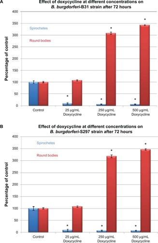
Figure 1B Susceptibility of the spirochete and round body forms of strain B31 (top panels) and strain S297 (bottom panels) of B. burgdorferi to different concentrations (between calculated MIC and MBC) of five antibiotics after 72-hour treatment measured by dark-field microscopy.
Note: *P values <0.05 indicates statistical significance compared with control.
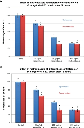
Figure 1C Susceptibility of the spirochete and round body forms of strain B31 (top panels) and strain S297 (bottom panels) of B. burgdorferi to different concentrations (between calculated MIC and MBC) of five antibiotics after 72-hour treatment measured by dark-field microscopy.
Note: *P values <0.05 indicates statistical significance compared with control.
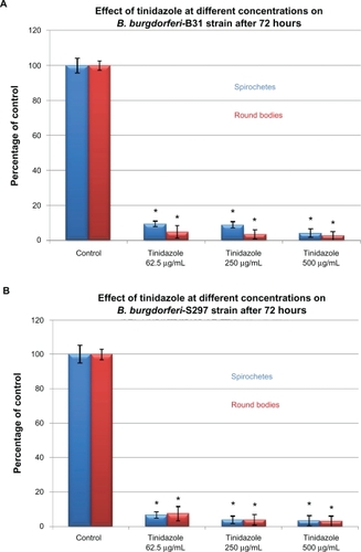
Figure 1D Susceptibility of the spirochete and round body forms of strain B31 (top panels) and strain S297 (bottom panels) of B. burgdorferi to different concentrations (between calculated MIC and MBC) of five antibiotics after 72-hour treatment measured by dark-field microscopy.
Note: *P values <0.05 indicates statistical significance compared with control.
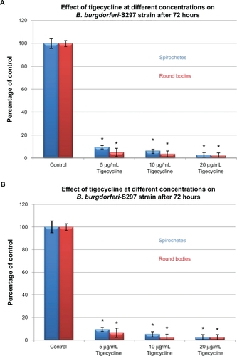
Figure 1E Susceptibility of the spirochete and round body forms of strain B31 (top panels) and strain S297 (bottom panels) of B. burgdorferi to different concentrations (between calculated MIC and MBC) of five antibiotics after 72-hour treatment measured by dark-field microscopy.
Note: *P values <0.05 indicates statistical significance compared with control.
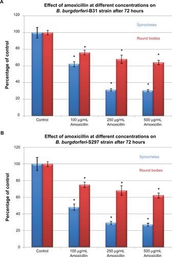
Figure 2A Susceptibility of the spirochete and round body forms of strain B31 (top panels) and strain S297 (bottom panels) of B. burgdorferi to the most effective concentrations of three antibiotics measured by dark-field microscopy. Tinidazole, metronidazole, and doxycycline effect on B. burgdorferi after 72-hour treatment.
Note: *P values <0.05 indicates statistical significance compared with control.
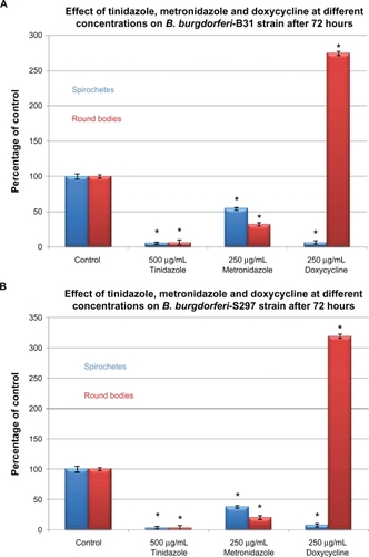
Figure 2B Susceptibility of the spirochete and round body forms of strain B31 (top panels) and strain S297 (bottom panels) of B. burgdorferi to the most effective concentrations of three antibiotics measured by dark-field microscopy. Tinidazole, metronidazole, and doxycycline effect on B. burgdorferi after 3 weeks of subculturing following 72-hour treatment.
Note: *P values <0.05 indicates statistical significance compared with control.
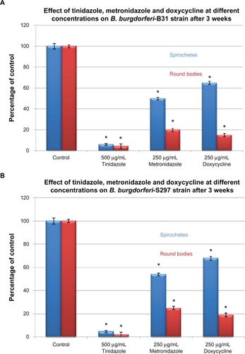
Figure 3A Evaluation of live/dead spirochete and round body forms of B. burgdorferi following treatment with five antibiotics measured by fluorescent microscopy using SYTO®9 green-fluorescent stain (live organisms) and propidium iodide red-fluorescent stain (dead organisms). Effect of doxycycline, tinidazole, tigecycline, metronidazole, and amoxicillin on spirochete forms of strain B31 (top panel) and strain S297 (bottom panel).
Notes: *P values calculated were <0.05 indicating statistical significance compared with control for live and dead spirochetes.
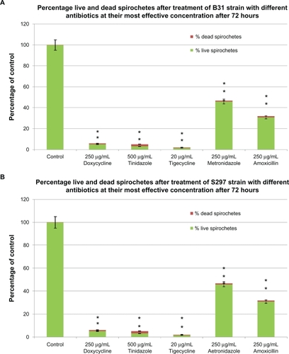
Figure 3B Evaluation of live/dead spirochete and round body forms of B. burgdorferi following treatment with five antibiotics measured by fluorescent microscopy using SYTO®9 green-fluorescent stain (live organisms) and propidium iodide red-fluorescent stain (dead organisms). Effect of doxycycline, tinidazole, tigecycline, metronidazole and amoxicillin on round body forms of strain B31 (top panel) and strain S297 (bottom panel).
Notes: *P values calculated were <0.05 indicating statistical significance compared with control for live and dead round bodies.
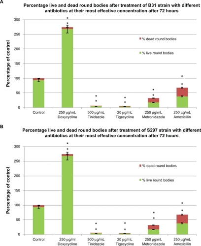
Figure 3C Evaluation of live/dead spirochete and round body forms of B. burgdorferi following treatment with five antibiotics measured by fluorescent microscopy using SYTO®9 green-fluorescent stain (live organisms) and propidium iodide red-fluorescent stain (dead organisms). Visualization of spirochete and round body forms of strain B31 following antibiotic treatment measured by dark field microscopy: (Ca) Control; (Cb) Doxycycline; (Cc) Tinidazole; (Cd) Metronidazole; (Ce) Tigecycline; (Cf) Amoxicillin.
Note: All images taken at 40× magnification.
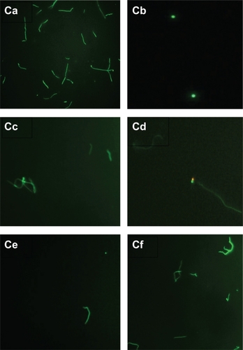
Figure 3D Evaluation of live/dead spirochete and round body forms of B. burgdorferi following treatment with five antibiotics measured by fluorescent microscopy using SYTO®9 green-fluorescent stain (live organisms) and propidium iodide red-fluorescent stain (dead organisms). Visualization of spirochete and round body forms of strain S297 following antibiotic treatment measured by dark field microscopy: (Da) Control; (Db) Doxycycline; (Dc) Tinidazole; (Dd) Metronidazole; (De) Tigecycline; (Df) Amoxicillin.
Note: All images taken at 40× magnification.
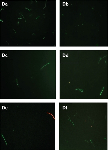
Figure 4A Evaluation of biofilm-like colonies of B. burgdorferi. Quantitative analysis of biofilm-like colonies of strain B31 (top panel) and strain S297 (bottom panel) measured by crystal violet staining technique.
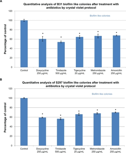
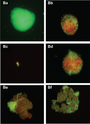
Figure 4B Evaluation of biofilm-like colonies of B. burgdorferi. Qualitative analysis of biofilm-like colonies of strain B31 measured by fluorescent microscopy using SYTO®9 green-fluorescent stain (live organisms) and propidium iodide red-fluorescent stain (dead organisms): (Ba) Control; (Bb) Doxycycline; (Bc) Tinidazole; (Bd) Tigecycline; (Be) Metronidazole; (Bf) Amoxicillin.
Note: All images taken at 40× magnification.