Figures & data
Figure 1 The content of N-linked and O-linked mannan in the cell wall of Candida albicans, Candida tropicalis, Candida guilliermondii, Candida krusei, and Candida auris.
Table 1 Cell wall analysis of C. albicans, C. tropicalis, C. guilliermondii, C. krusei, and C. auris
Notes: Yeast cells were treated either with endoglycosidase H or b-eliminated to trim N-linked mannans or O-linked mannans, respectively. The released oligosaccharides were saved and used to measure the mannose content by HPAEC-PAD. Data are mean ± SD of three independent experiments performed in duplicates. *P<0.05, when compared with mannans from the other species analyzed.
Abbreviation: HPAEC-PAD, high-performance anion-exchange chromatography coupled to pulsed amperometric detection.
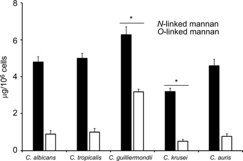
Table 1 Cell wall analysis of C. albicans, C. tropicalis, C. guilliermondii, C. krusei, and C. auris
Figure 2 Fluorescent labeling of chitin and b1,3-glucan in the cell wall of Candida albicans, Candida tropicalis, Candida guilliermondii, Candida krusei, and Candida auris.
Notes: Live or heat-killed (HK) yeast cells were incubated with either fluorescein isothiocyanate-wheat germ agglutinin conjugate (closed bars, labels chitin) or IgG Fc-Dectin-1 chimera (open bars, labels b1,3-glucan) as described in the “Materials and methods” section, inspected under fluorescence microscopy, and the fluorescence associated to 300 individual cells was recorded. *P<0.05, when compared with cells under the same treatment.
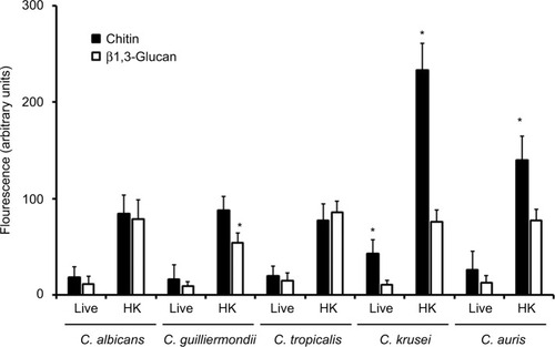
Figure 3 Stimulation of cytokine production by Candida albicans, Candida tropicalis, Candida guilliermondii, Candida krusei, and Candida auris.
Notes: Human PBMCs were coincubated for 24 hours with live yeast cells, and then the supernatant was collected and used to quantify the cytokine levels. *P<0.05, when compared with the cytokine level stimulated by C. albicans cells.
Abbreviations: PBMCs, peripheral blood mononuclear cells; TNFα, tumor necrosis factor alpha.
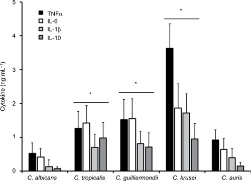
Figure 4 Stimulation of cytokine production by heat-killed and b-eliminated cells from Candida albicans, Candida tropicalis, Candida guilliermondii, Candida krusei, and Candida auris.
Notes: Yeast cells were heat-killed (HK), b-eliminated, or subjected to both treatments before being coincubated with human PBMCs for 24 hours. The supernatants of interactions were collected and used to quantify the cytokine levels. * P<0.05, when compared with the cytokine level stimulated by C. albicans cells under the same treatment. Abbreviations: PBMCs, peripheral blood mononuclear cells; TNFα, tumor necrosis factor alpha; C. alb, Candida albicans; C. tro, Candida tropicalis; C. gui, Candida guilliermondii; C. kru, Candida krusei; C. aur, Candida auris.
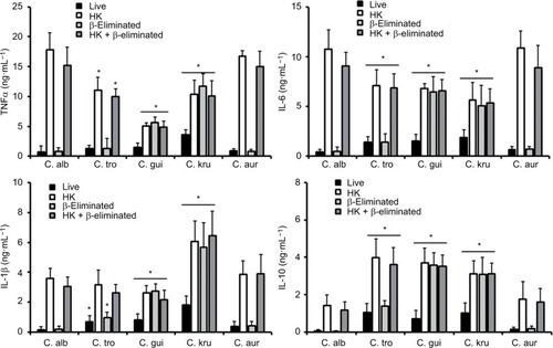
Table 2 Effect of laminarin on the ability of C. albicans, C. tropicalis, C. guilliermondii, C. krusei, and C. auris to stimulate cytokine production
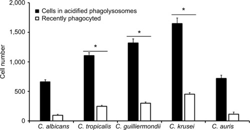
Figure 5 Phagocytosis of Candida albicans, Candida tropicalis, Candida guilliermondii, Candida krusei, and Candida auris by human monocyte-derived macrophages.
Notes: Acridine orange-labeled yeast cells were incubated with the human cells at an MOI ratio of 1:6 for 2.5 hours at 37°C under a CO2 atmosphere. Then, macrophages were gated by FACS system and 50,000 cells were counted/sample. Results represent macrophages interacting with at least one green fluorescent cell (recently phagocyted), and those associated with red fluorescence that were classified as macrophages with yeast cells within acidified phagolysosomes. The data represent the mean ± SD of three independent biological replicates performed in duplicate. * P<0.05, when compared with C. albicans cells.
Abbreviations: MOI, multiplicity of infection; FACS, fluorescence-activated cell sorter.