Figures & data
Table 1 Characteristics of the patients
Figure 1 Clinical features of mycobacterium fortuitum endophthalmitis. (A) Biomicroscopic examination identified conjunctival congestion (represented by the white arrow) and a distorted pupil obscured by hypopyon (the pupillary margin hidden in the circle). (B) Biomicroscopic examination identified a severe fibrinous reaction (the fibrinous tissues in the circle) and hypopyon in front of the intraocular lens (represented by the black arrow). (C and D) The vitreous and fundus have a yellow and white purulent exudate. (E) A B-scan ocular ultrasound identified vitreous opacification.
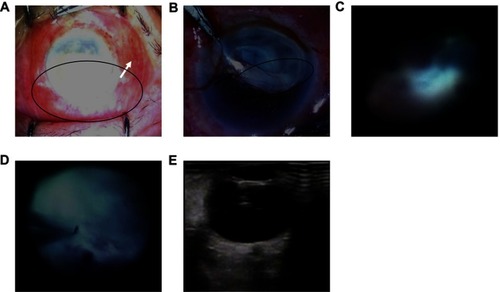
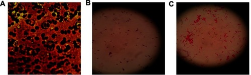
Figure 2 Diagnosis of Mycobacterium fortuitum endophthalmitis. (A) Microscopic examination of the anterior chamber fibrin membrane sample isolated after vitrectomy. (B) Gram stain of the anterior chamber fibrin membrane sample isolated after vitrectomy. (C) Acid-fast stain of the anterior chamber fibrin membrane sample detected acid-fast bacilli (red stain) that were identified as Mycobacterium fortuitum.